

A mast cell tumor in cats is a cancer of immune cells called mast cells. A single, low-grade skin tumor is often cured by surgery, and many cats live a normal lifespan. Tumors in the spleen or intestines are more serious, with survival typically measured in months even with treatment.
A mast cell tumor in cats is a cancer that arises from mast cells, a normal part of the immune system that stores histamine and heparin. It’s one of the most common feline skin tumors, but it can also develop inside organs such as the spleen and intestines. If your veterinarian has found a suspicious lump or diagnosed a mast cell tumor, you’ll likely want to know how serious it is and what comes next. For most cats with a single, low-grade skin tumor, the outlook is highly favorable, and surgery often cures it. Tumors in internal organs such as the spleen or intestines have a more guarded prognosis, but treatment options and meaningful survival times are still possible. Diagnostic testing, tumor grading, and location help determine whether the disease is confined to the skin or affects the body more broadly.
Mast Cell Tumors in Cats: What They Are and the Short Answer on Seriousness
A mast cell tumor in cats is a neoplasm of specialized immune cells that help manage inflammatory responses. Cutaneous tumors, which occur on the skin, often behave benignly or are easy to manage. Visceral tumors require careful veterinary oversight. An individual tumor’s behavior depends on its location, cellular grade, and specific subtype. Its appearance alone can’t reveal how aggressive it is, so a visual exam can’t provide a definitive answer.
What a mast cell tumor on a cat looks like
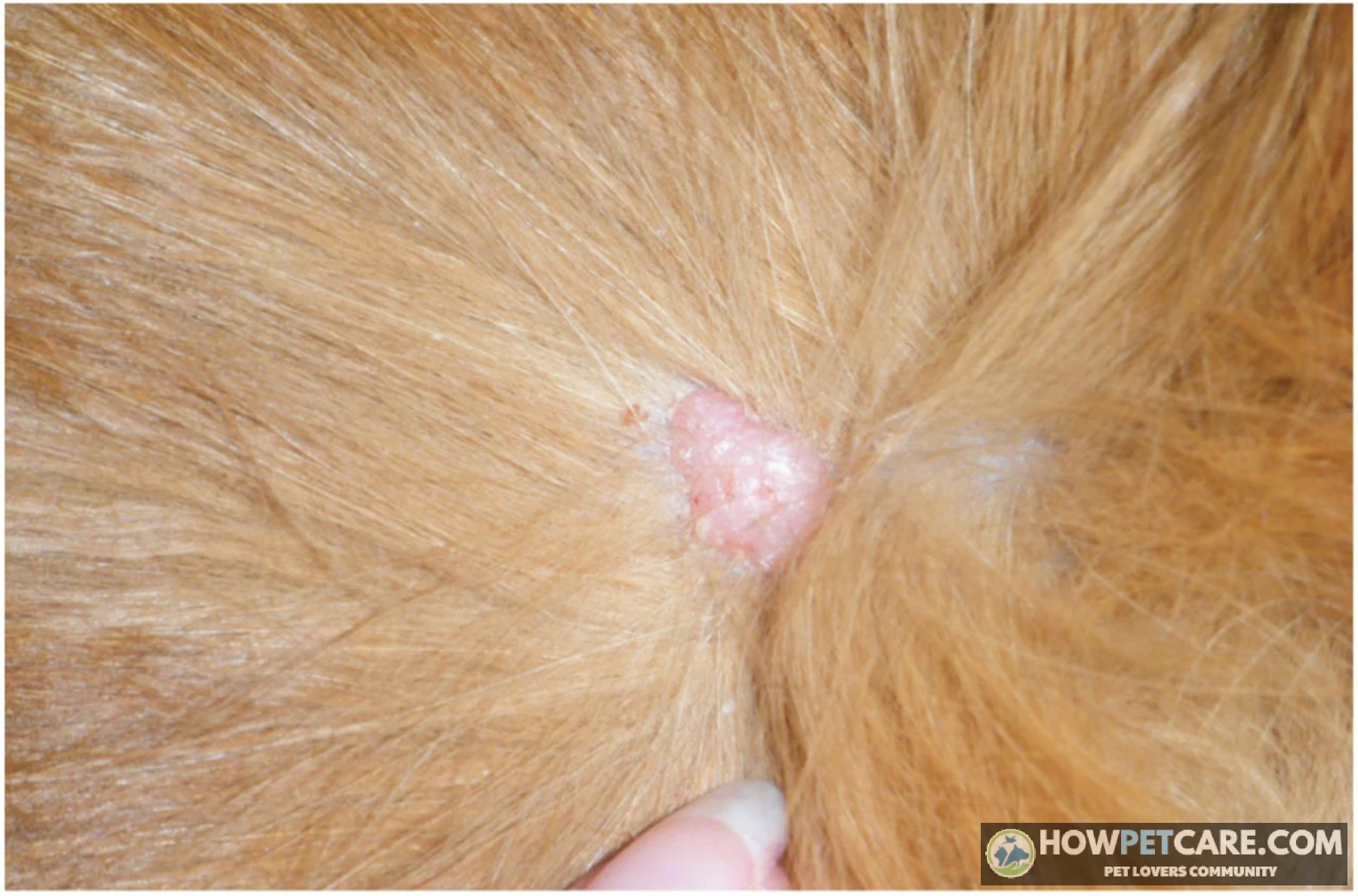
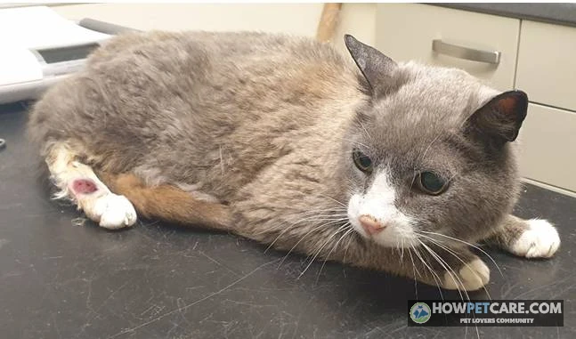
On the skin, a mast cell tumor typically appears as a firm, raised, hairless, or pink nodule measuring between 0.5 and 1 centimeters across. These lesions commonly occur on the head, neck, trunk, or limbs. They may appear as a single mass or in multiple clusters and often cause itching or surface ulceration. Early on, they can look like a harmless pimple, a small insect bite, or a benign skin tag.
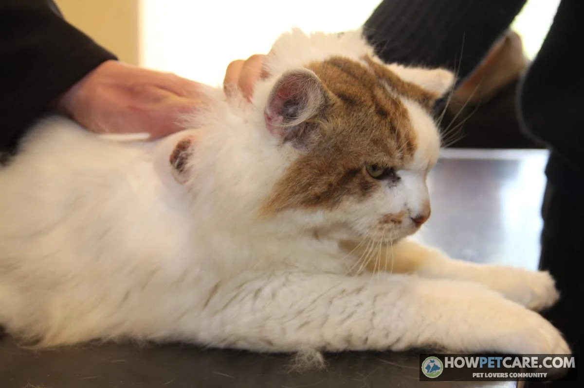
Don’t squeeze, massage, or vigorously poke any lump on your cat’s skin. Handling a mast cell tumor can trigger degranulation—the sudden release of stored histamine and heparin from the cells. This can cause local swelling, redness, and hives, as well as systemic problems such as nausea or stomach upset.
Why you can’t tell a mast cell tumor from other skin tumors by sight
Many feline skin conditions look alike, from benign cysts to other cancers such as basal cell tumors or fibrosarcomas. A veterinarian can’t identify or grade a mass just by looking at it or feeling it. A diagnosis requires examination under a microscope.
Cutaneous vs. Splenic vs. Intestinal: How Location Changes Severity
A mast cell tumor’s location has a major influence on its behavior, symptoms, and treatment. Feline mast cell disease generally falls into three main anatomical categories: cutaneous (skin), splenic (visceral), and intestinal.
Cutaneous mast cell tumors (including ear, head, and mouth)
Cutaneous mast cell tumors are the most common form and the second most frequent skin tumor in cats. They typically appear as firm skin lumps that may itch or ulcerate. They often develop on the head, neck, and ears, including the pinna and the base of the earflap. Mast cell tumors can also occur inside the mouth, although this is rare; there, they may bleed or interfere with eating. A specialized variant called the histiocytic subtype occurs mainly in young cats, particularly Siamese and related Oriental breeds, and often appears as multiple nodules. Unlike standard mastocytic tumors, the histiocytic form can sometimes regress on its own without aggressive intervention.
Splenic and visceral mast cell tumors
Splenic mast cell tumors are the most common tumor type found in the feline spleen and a primary form of visceral disease. When mast cells infiltrate internal organs such as the spleen or liver, symptoms are often systemic and non-specific. Owners may notice chronic vomiting, decreased appetite, progressive weight loss, lethargy, pale mucous membranes, or an enlarged, fluid-filled abdomen. Affected cats frequently develop secondary stomach ulcers because mast cells continuously release histamine into the bloodstream. Splenectomy, or removal of the spleen, is the standard treatment for splenic disease.
Intestinal mast cell tumors
Intestinal mast cell tumors are less common but are among the more aggressive forms of feline mast cell disease. Symptoms include severe gastrointestinal upset, with persistent vomiting, chronic diarrhea, profound weight loss, and anorexia. These tumors are more likely to metastasize to nearby lymph nodes and other organs, making the outlook more guarded.
Mast cell tumors in lymph nodes
Lymph nodes can become involved when a primary skin or visceral tumor spreads or, rarely, as the primary site of origin. If lymph nodes near a skin tumor or within the abdomen are enlarged, a veterinarian may recommend fine-needle aspiration to check for tumor cells that have spread.
 You might be wonderingHairballs in cats treatmentHairballs in Cats Treatment: A Step-by-Step Home ProtocolRead the answer
How Vets Diagnose a Mast Cell Tumor: FNA, Biopsy, and Staging
A veterinary workup for feline mast cell disease can begin with a quick preliminary test and progress to pathology and whole-body staging.
Fine needle aspiration: what it can and can’t tell you
For a fine needle aspiration (FNA), the veterinarian inserts a small needle into the lump to collect cells, which are stained and examined under a microscope. The procedure is quick, usually requires no sedation, and costs between $100 and $250. An FNA can reliably identify mast cells, but it can’t always establish the exact histological grade or predict how the tumor will behave.

Biopsy and histopathology
Histopathology is the laboratory examination of a tissue biopsy, taken either by removing the entire mass or by collecting a wedge sample. It’s the definitive diagnostic step, revealing the tumor’s subtype, cellular differentiation, mitotic index, and whether the surgical margins are clean. For many small skin tumors, the entire mass is removed surgically and sent for histopathology.
Staging: ultrasound, bloodwork, and lymph node checks
A complete staging evaluation is recommended when a cat has a visceral tumor, a poorly differentiated cutaneous tumor, or multiple masses. Staging typically includes complete blood counts and chemistry panels to screen for anemia and circulating mast cells, an abdominal ultrasound to image the spleen and liver, and fine-needle aspirations of nearby lymph nodes. Chest X-rays are also performed, although spread to the lungs is less common in cats than in dogs.
How to Read Your Cat’s Pathology Report: Grade, Subtype, and Mitotic Index Explained
Your cat’s pathology report contains specialized terms that help guide prognosis and treatment. Feline reports use criteria distinct from canine grading systems.
| Report term | Plain-language meaning | What it suggests for prognosis | Question to ask your vet |
|---|---|---|---|
| Subtype: mastocytic | The common form — tumor made of mast cells | Behavior then depends on differentiation (below) | "Is this the mastocytic or histiocytic form?" |
| Subtype: histiocytic | A different cell type, seen mostly in young cats and Siamese, often with multiple nodules | Can behave mildly and sometimes regresses on its own | "Does this change whether we need to treat right away?" |
| Well-differentiated | Cells look close to normal mast cells | Low-grade behavior; surgery is often curative | "Is complete removal likely to be the end of this?" |
| Poorly differentiated / pleomorphic | Cells look abnormal and disorganized | Higher risk of recurrence and spread | "Should we see an oncologist and stage the rest of the body?" |
| Mitotic index / mitotic count | How many cells were dividing per 10 high-power microscope fields | Low is favorable; counts above roughly 5/10 hpf in feline studies link to shorter survival | "What was the exact count, and what does it change about treatment?" |
| Margins: complete / clean | The tumor was removed with healthy tissue all around it | Recurrence risk is low | "How wide were the margins?" |
| Margins: incomplete / dirty | Tumor cells reached the edge of the removed tissue | Regrowth is possible; options are monitoring, re-surgery, or radiation | "Do we monitor, re-operate, or radiate — and why?" |
| Comments / description | The pathologist’s free-text notes: ulceration, depth, eosinophils | Context for the findings above | "Is there anything in the comments that worries you?" |
Well-differentiated vs. poorly differentiated
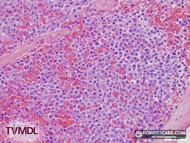
Well-differentiated mast cell tumors contain cells that closely resemble normal, healthy mast cells. These masses typically behave in a benign or low-grade fashion, and complete surgical removal often cures them. Poorly differentiated or pleomorphic tumors, by contrast, have disorganized, highly atypical cells and are more likely to recur locally or spread through the body.
Histiocytic subtype
The histiocytic subtype has distinct round cells and occurs mainly in young cats, particularly Siamese breeds. These tumors often appear as multiple skin nodules that can regress on their own over several weeks or months without medical treatment.
Mitotic index and surgical margins
The mitotic index measures how often cells are actively dividing across high-power microscope fields. Published feline oncology studies link a low mitotic count with a favorable long-term outlook. Counts exceeding approximately 5 mitoses per 10 high-power fields are associated with more aggressive tumor behavior. Surgical margins describe the border of healthy tissue around the removed tumor. Clean margins mean the cancer was fully removed; dirty margins indicate that tumor cells extend to the edge of the tissue sample.
How Long Can a Cat Live With A Mast Cell Tumor? Survival Times by Type, Grade, and Treatment
The outlook differs considerably between a tumor confined to the skin and disease involving internal organs such as the spleen. Cats whose low-grade skin tumors are surgically removed often live a normal lifespan. With systemic or visceral disease, survival is measured in months.
| Tumor location / type | Differentiation or grade | Treatment | Typical survival or outcome (published median/range) | Notes for owners |
|---|---|---|---|---|
| Cutaneous (skin), single tumor | Well-differentiated, low mitotic count | Surgical removal with clean margins | Often cured; many cats live a normal lifespan (years). Local recurrence is uncommon | The best-case scenario — most feline skin MCTs fall here |
| Cutaneous, single tumor | Well-differentiated, incompletely removed | Monitoring, second surgery, or radiation | Still frequently years; recurrence is the main risk, not spread | An incomplete margin does not automatically mean relapse — ask about monitoring vs. re-treatment |
| Cutaneous | Poorly differentiated or high mitotic count (about >5 per 10 high-power fields) | Surgery plus chemotherapy and/or radiation | Roughly months to ~2 years in published high-grade groups | Oncologist referral strongly recommended; response varies widely |
| Histiocytic subtype (often young cats, Siamese, multiple nodules) | N/A — classified by subtype, not standard grade | Sometimes monitoring alone; surgery if needed | Variable; some nodules regress on their own | Do not assume the worst — ask whether your cat’s report says ‘histiocytic’ |
| Splenic (visceral) | N/A — spleen disease is treated as systemic | Splenectomy, sometimes plus chemotherapy | Median roughly 12–19 months (commonly cited ~14 months); some cats live much longer | The spleen is usually removed even if the cat feels fine — it can rupture and bleed |
| Splenic | N/A | No surgery | Typically weeks to a few months | Progression and internal bleeding are the main risks |
| Intestinal | Usually behaves aggressively | Surgery plus chemotherapy where possible | Typically months (often under 6); a minority respond longer | The most guarded prognosis; ask honestly about quality-of-life goals |
| Metastatic (lymph nodes, liver, blood involvement) | Any | Chemotherapy, targeted drugs (Palladia), palliative care | Weeks to months; occasionally longer responders | Focus shifts to comfort and slowing progression |
What happens if you choose not to treat
An untreated low-grade skin mast cell tumor may stay the same size for a long time or grow slowly. Untreated visceral or splenic disease, however, progresses steadily and can lead to worsening anemia, abdominal discomfort, internal bleeding, and systemic decline. When curative treatment isn’t feasible, palliative supportive care focuses on comfort and control of histamine-related symptoms.
Treatment Options for Feline Mast Cell Tumors: Surgery, Chemotherapy, Palladia, and Supportive Care (With Costs)
Treatment can be local or systemic, depending on the diagnosis and your cat’s overall health.
Surgery and radiation
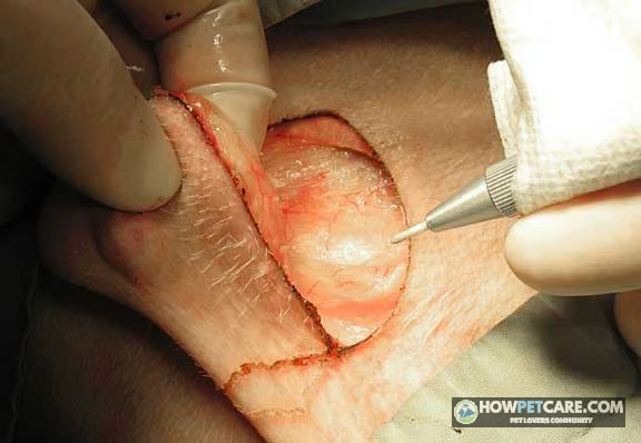
Surgery is the primary treatment for most cutaneous mast cell tumors. It typically requires margins of 1 to 2 centimeters where the anatomy allows. Skin tumor removal costs range from $800 to $4,000, while a splenectomy for visceral disease generally costs between $2,000 and $6,000. Radiation therapy is used for incompletely removed tumors or masses in delicate areas such as the face, ear, or limbs, where wide margins aren’t possible. Definitive radiation protocols cost between $10,000 and $12,000, while palliative radiation ranges from $2,500 to $6,000.
Chemotherapy: lomustine, vinblastine, and chlorambucil
Chemotherapy drugs such as lomustine (CCNU), vinblastine, and chlorambucil are prescribed for high-grade, recurrent, or visceral mast cell tumors, often alongside corticosteroids such as prednisolone. They aim to slow tumor progression and manage disease that has spread. Each chemotherapy session typically costs between $600 and $900, including the monitoring bloodwork needed to check white blood cell counts and organ function.
Palladia and other targeted drugs
Tyrosine kinase inhibitors such as toceranib (Palladia) and masitinib are targeted oral therapies used off-label in cats with refractory or aggressive mast cell disease. These medications cost from $600 to $800 per month.
Prednisolone, H2 blockers, and antihistamines
Corticosteroids such as prednisolone are often used to reduce tumor inflammation and shrink masses. H2-blockers such as famotidine and standard antihistamines are also given to reduce the systemic effects of histamine release, prevent gastric ulceration, and reduce nausea.
| Service or treatment | What it involves | Typical cost range (USD) | When it’s used |
|---|---|---|---|
| Fine needle aspiration (FNA) + cytology | Needle sample of the lump read under a microscope | $100–$250 | First step for any new lump; usually no sedation |
| Histopathology (biopsy) | Pathologist grades the removed tissue | $150–$400 on top of surgery | Every removed lump should be sent in |
| Bloodwork (CBC, chemistry) | Screens for anemia and organ health | $150–$300 | Before anesthesia and for staging |
| Abdominal ultrasound | Images spleen, liver, lymph nodes | $300–$600 | Staging visceral or high-grade disease |
| Chest X-rays | Checks lungs (less central in cats than dogs) | $150–$300 | If staging is recommended |
| Skin tumor removal (excision) | Surgery with ~1–2 cm margins plus sutures | $800–$4,000 typical; up to ~$8,000 for difficult sites | First-line for cutaneous tumors |
| Splenectomy | Removal of the spleen under general anesthesia | $2,000–$6,000 | Standard treatment for splenic MCT |
| Chemotherapy (lomustine, vinblastine, chlorambucil) | Per-session drug cost plus monitoring bloodwork | ~$600–$900 per session | High-grade, recurrent, or visceral disease |
| Palladia (toceranib) / masitinib | Oral targeted drug, off-label in cats | ~$600–$800 per month | Aggressive or metastatic disease |
| Radiation therapy | Multiple sessions over weeks | $2,500–$12,000 by protocol | Incomplete margins or hard-to-reach sites (face, ear, limbs) |
| Oncologist consultation | Specialist review of report and options | $200–$400 | Before any chemo/radiation decision |
What to Do After a Mast Cell Tumor Diagnosis: A Step-by-Step Decision Path
A cancer diagnosis can feel overwhelming. Working through the findings with your veterinarian can help you decide what comes next.
-
Step 1: Confirm diagnosis and location
Confirm whether the mass is a single skin nodule, multiple cutaneous lesions, or an internal finding in the spleen or intestines.
-
Step 2: Review pathology and margins
Get the complete pathology report to identify the tumor’s exact subtype, mitotic index, level of cell differentiation, and margin status.
-
Step 3: Complete staging if indicated
Complete bloodwork, abdominal ultrasound, or lymph node aspirations if the tumor is poorly differentiated, visceral, or recurrent.
-
Step 4: Select the treatment path
Choose between surgical removal alone, combination therapy, radiation, or medical management based on your cat’s health and staging results.
When to see a veterinary oncologist
A consultation with a board-certified veterinary oncologist (costing $200–$400) is strongly recommended if your cat’s pathology report shows a poorly differentiated tumor, a high mitotic index, visceral or intestinal involvement, regional lymph node spread, or a difficult anatomical location where surgery is challenging. Oncologists have specialized expertise in chemotherapy protocols and targeted treatments.

Seek immediate veterinary care if your cat develops sudden abdominal distention, pale gums, rapid breathing, collapse, repeated vomiting, or black, tarry stools. These signs can indicate internal bleeding or severe systemic histamine reactions.
Home Lump Checks and Life After Mast Cell Tumor Surgery
Checking your cat at home can help you catch new or recurring masses early and support their quality of life.
-
1. Prepare your tracking tools
Gather your phone camera, a small ruler or coin for scale, and a dedicated notebook or digital log.
-
2. Inspect the head and neck
Using flat fingers, feel gently behind and under the ears and along the jawline, chin, and cheeks.
-
3. Check shoulders and front legs
Stroke down the neck, check the armpits, and run your fingers down each front leg to the paws.
-
4. Examine the torso and belly
Gently feel the chest, ribs, and abdomen, as well as the groin where the hind legs meet the body.
-
5. Document and report
Record the location, size, and date of any new finding, and schedule a veterinary evaluation promptly.
Surgery recovery: cone, activity, and incision care
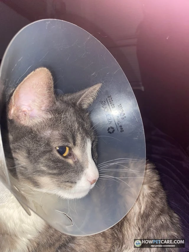
After skin tumor removal, cats must wear an Elizabethan collar or recovery suit for 10 to 14 days, until the sutures are removed. Activity must be strictly restricted: no jumping on furniture, running, or rough play. Check the incision daily for swelling, excessive redness, discharge, or suture breakdown.
Monitoring for recurrence or new tumors
Cats that develop one mast cell tumor are more likely to develop new masses elsewhere on the body. A monthly home lump check and scheduled veterinary rechecks every three to six months help identify future changes as early as possible, when they’re most treatable.
Your Next Vet Visit: Questions to Ask and a Checklist to Bring
Preparing for the visit can help you make the most of your time with your veterinarian or veterinary oncologist. Bring the complete pathology report, all bloodwork and imaging results, a clear log of any findings at home, and a list of current medications.
- What is the exact subtype and histological grade of my cat’s tumor?
- What was the mitotic count, and were the surgical margins completely clean?
- Is whole-body staging necessary, or can we proceed directly with localized treatment?
- Should we consult a veterinary oncologist for advanced therapy options?
- What itemized cost estimates apply to the next phases of care?
Use your cat’s specific diagnosis, your financial comfort level, and their daily comfort to guide the next decision with your veterinary team.
 You might be wonderingWorms in catsWorms in Cats: Symptoms, Types, Treatment & When to See a VetRead the answer
FAQ
Are certain cat breeds more likely to get mast cell tumors?
Siamese cats and related Oriental breeds are overrepresented, especially for the histiocytic form that appears in younger cats. Mast cell tumors can occur in any breed, and most affected cats are middle-aged or older.
Is a mast cell tumor in a cat painful?
Most skin tumors are not painful, though they can be itchy, red, or ulcerated. Visceral forms can cause nausea, poor appetite, and abdominal discomfort from histamine release and organ involvement.
Can a cat’s mast cell tumor go away on its own?
Some histiocytic-type tumors in young cats have been reported to shrink or disappear without treatment, but this is not something to count on. Any lump should still be sampled and monitored by a vet.
Is a mast cell tumor in cats contagious to other pets or people?
No. Mast cell tumors are not infectious and cannot be passed to other pets or to people.
Can I give my cat Benadryl for a mast cell tumor?
Do not give any human antihistamine or stomach medication without your vet’s instruction. Vets sometimes prescribe antihistamines or famotidine to cats with mast cell disease, but the product and dose must be chosen for cats.