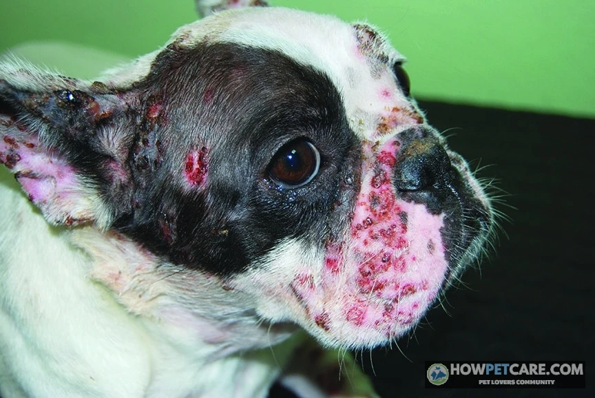

Pemphigus foliaceus (PF) is the most common autoimmune skin disease diagnosed in dogs. The immune system attacks the connections between skin cells, causing fragile pustules, crusts, and hair loss—usually starting on the face and ears. It isn’t contagious, and most dogs respond to immunosuppressive medication over months.
Pemphigus Foliaceus in Dogs: What It Is and What to Expect
Pemphigus foliaceus in dogs is an autoimmune skin disease that can cause fragile pustules, thick crusts, and hair loss, often starting on the face and ears. It isn’t contagious to people or other dogs. A veterinarian can usually manage it with medication and monitoring, though treatment may take months and some dogs need ongoing care. PF is the most commonly diagnosed autoimmune skin disease in dogs.
If your dog has recurring facial crusts or sore footpads, book a veterinary exam instead of trying to diagnose the problem from appearance alone. Recent veterinary studies put complete remission at approximately 52% of affected dogs, though results differ between studies and individual dogs.
How the autoimmune attack works, in plain English
Normally, structures called desmosomes help hold skin cells together. In canine pemphigus foliaceus, the immune system produces antibodies that interfere with these connections. That separation of skin cells is called acantholysis; it helps create pustules and weakens the skin barrier. Adhesion proteins such as desmoglein have been studied as targets, though the immune response isn’t identical in every dog.
Is pemphigus foliaceus contagious to other dogs or people?
No. PF itself is an immune system disorder, not an infection that spreads between dogs or to people. However, an infection that resembles PF—or an infection occurring alongside it—may require separate precautions. Your veterinarian can advise you while test results are pending.
Early Signs of Pemphigus Foliaceus in Dogs: A Lesion-Recognition Checklist
Where PF lesions show up first
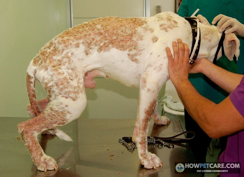
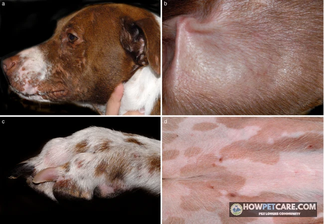
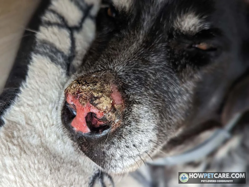
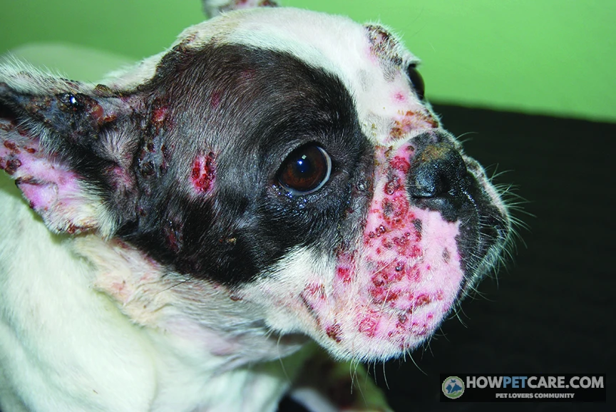
Early signs of pemphigus foliaceus in dogs often involve the bridge of the nose, the skin around the eyes, and the ear flaps. Some dogs develop thickened, cracked, or crusted footpads or inflamed nail beds. Lesions can later spread to the trunk and other areas.
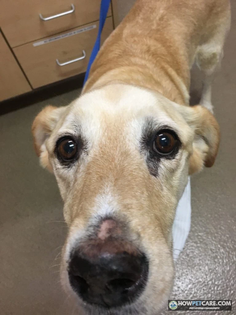
Itching varies: a dog may scratch a great deal or show little itch. Eroded skin and cracked pads can be painful, and limping may be the first sign an owner notices. More widespread disease can bring fever, lethargy, poor appetite, swollen lymph nodes, or limb swelling.
From pustule to crust: how a lesion changes
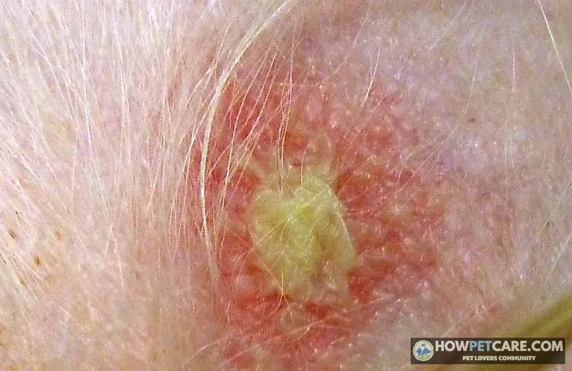
A spot may begin as redness or a small bump, then form a pus-filled blister called a pustule. These pustules are fragile and can rupture quickly, so owners often see yellow-tan or honey-colored crusts rather than intact blisters. Scaling, superficial raw areas, and hair loss may follow.
PF flare vs. ordinary scab vs. bacterial skin infection
Use this checklist to decide what to tell your veterinarian—not to identify PF at home:
- Location: Are crusts or pustules appearing on the nose, around the eyes, on the ears, or around the feet?
- Distribution: Are similar areas affected on both sides of the face or body?
- Appearance: Are there recurrent, sometimes layered yellow-tan crusts, with hair loss or raw skin beneath them?
- Pattern: Do lesions return in the same places or appear during a medication taper?
- Response: Has the problem persisted despite a complete course of antibiotics prescribed for a suspected infection?
- Whole-dog signs: Is your dog limping, less energetic, feverish, or eating poorly?
An ordinary scab often follows a recognizable injury and heals without repeatedly producing new lesions. Bacterial skin infection can also cause pustules and crusts, however, and may occur at the same time as PF. Symmetry or poor antibiotic response raises a question; neither proves the answer. Do not remove crusts to check whether they return.
Recurrent facial crusting, new pustules, or painful feet warrant a veterinary visit even if your dog otherwise seems well. Ask whether cytology and a skin biopsy are appropriate. This article can’t diagnose your dog.
Which Dogs Get Pemphigus Foliaceus? Breeds, Age and Triggers
PF can affect dogs of different ages and breeds, though it is often diagnosed in adulthood. Breeds reported as predisposed include Akitas, Chow Chows, Doberman Pinschers, Dachshunds, Newfoundlands, and Bearded Collies. These associations do not mean a dog of one of these breeds will develop PF, and reliable breed-specific risk percentages are not established here.
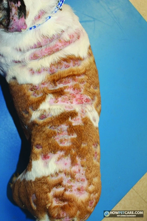
In many dogs, no specific cause is identified. Some cases have been associated with medications or have developed in dogs with longstanding skin inflammation. Give your veterinarian a complete list of prescriptions, supplements, and parasite preventives, including when each was started. Do not stop a prescribed drug without discussing it first.
Sunlight can aggravate lesions in some dogs, particularly on exposed facial skin. An association with a trigger does not mean an owner caused the disease.
How Vets Diagnose Pemphigus Foliaceus and Rule Out Look-Alikes
The diagnostic workup, step by step
A veterinarian considers the appearance and distribution of lesions alongside test results. The process may include:
- History and examination. Your vet will ask when the lesions began, which areas were affected first, what medicines your dog takes, and whether earlier treatments helped.
- Cytology. A sample from a pustule or beneath a crust is examined on a slide. Rounded, separated skin cells called acantholytic keratinocytes can support suspicion of PF. Cytology can also reveal signs of infection, but it doesn’t establish PF on its own.
- Tests for mimics. Skin scrapings can look for mites; fungal testing can investigate ringworm; and additional testing may be needed for bacterial infection. Infection and PF can coexist.
- Skin biopsy. A vet takes small tissue samples, often from more than one fresh lesion, for examination by a pathologist. Biopsy is the key confirmatory test when interpreted with the examination and other results. Samples may be collected using local anesthesia, sedation, or another plan suited to the dog.
- Baseline laboratory tests. Bloodwork and often urinalysis help assess your dog’s health before immunosuppressive therapy and provide a comparison for later monitoring.
Ask the clinic how to prepare for sampling. Do not scrub lesions or apply new products beforehand unless directed. If your dog already takes a steroid, tell the vet: medication can affect biopsy interpretation, but do not stop it on your own.
Pemphigus foliaceus vs. vulgaris, erythematosus and vegetans
| Variant | Typical lesion locations | Lesion type | Relative frequency and severity | How it is confirmed |
|---|---|---|---|---|
| Foliaceus | Face, ears, feet; sometimes widespread skin | Fragile pustules followed by crusts and erosions | Most common canine pemphigus form; severity varies | Examination, cytology, and biopsy |
| Erythematosus | Mainly face and nose | Localized crusting and sometimes pigment loss | Often milder and more localized | Biopsy and exclusion of similar diseases |
| Vulgaris | Mouth, lips, other mucocutaneous junctions, and sometimes skin | Deeper erosions or ulcers | Rare and often more severe; mouth pain can be significant | Biopsy showing a deeper pattern of skin-cell separation |
| Vegetans | Skin folds and other affected skin | Raised, wart-like lesions that may erode | Rare | Biopsy interpreted with the clinical appearance |
PF is not generally described as turning into pemphigus vulgaris. Tell your vet promptly if your dog develops mouth sores, drooling, or difficulty eating; those signs need their own assessment.
Diseases that look like PF
Other possible explanations for crusting include bacterial pyoderma, ringworm, demodectic mange, discoid lupus, zinc-responsive dermatosis, drug eruptions, and, less commonly, cutaneous lymphoma. Travel history can matter when considering infections such as leishmaniasis. Appearance alone cannot reliably distinguish these conditions, and a dog can have more than one skin problem at once.
Pemphigus Foliaceus Treatment in Dogs: Drugs, Tapering and the 4–12 Week Timeline
Treatment aims first to stop new lesions and relieve discomfort, then to find the lowest effective treatment plan. The choice depends on how much skin is affected, other health conditions, test results, and how your dog tolerates medication. Your veterinarian—not an online guide—must determine doses.
| Drug | Role in treatment | Typical onset of effect | Main side effects to watch | Typical monitoring |
|---|---|---|---|---|
| Prednisone or prednisolone | Systemic corticosteroid commonly used to control widespread disease | Improvement may begin in the first few weeks | Thirst, urination, hunger, panting, muscle weakness, and infections | Exams, bloodwork, and sometimes urine testing |
| Azathioprine | Immunosuppressant often added in dogs to help reduce steroid exposure | Can take several weeks | Vomiting, diarrhea, lethargy, infection, or unusual bruising | Complete blood count and liver tests |
| Cyclosporine | Alternative immunosuppressant when appropriate | Response varies and may take weeks | Digestive upset, gum overgrowth, and infections | Exams and laboratory testing as directed |
| Chlorambucil | Another immunosuppressant a specialist may consider for difficult cases | Response varies | Digestive upset and reduced blood-cell counts | Regular blood counts and other tests as directed |
| Topical tacrolimus | Local immune-modulating treatment sometimes used for limited lesions | Response varies | Local irritation; prevent licking | Skin rechecks and review of application safety |
For a mild, localized case, a veterinarian may consider a topical corticosteroid or tacrolimus, sometimes without systemic medication. More extensive disease commonly requires systemic treatment. Azathioprine is a familiar steroid-sparing option in dogs; chlorambucil may be considered when other approaches are unsuitable or unsuccessful. A steroid-sparing drug doesn’t mean the steroid can be stopped immediately.
What the first 12 weeks usually look like
- Weeks 0–4: The first plan focuses on stopping new lesions, easing discomfort, and treating any confirmed infection. Take photographs so changes are easier to judge.
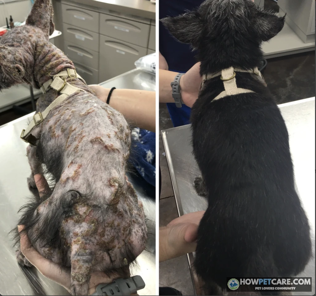
- Weeks 4–12: Some dogs improve in the first few weeks; initial clinical remission may take roughly 4–12 weeks of intensive treatment. That range isn’t a promise, and some dogs need longer or a change in plan.
- After control: Once the vet is satisfied that new lesions are controlled, the discussion turns to gradual tapering and the least medication needed to maintain comfort.

Healing crusts can remain visible after new pustules have stopped forming. Conversely, a few new lesions during a taper may be important even if most of the skin looks better. Share both observations with the vet.
Tapering and lab monitoring: what a typical schedule looks like
A taper usually takes place gradually over months, with adjustments based on the dog’s skin and side effects. Rechecks and blood tests are generally closer together when treatment begins or changes, then may be spaced out once the dog is stable. Depending on the medication and the dog, monitoring can include a complete blood count, liver and other chemistry values, urinalysis, and sometimes urine culture.
Ask your vet for the exact dates and tests appropriate to your dog’s prescription. Never abruptly stop or reduce a steroid yourself: a sudden change can cause a flare and may also be dangerous because prolonged steroid treatment can suppress the body’s normal adrenal function.
Managing steroid side effects
Increased thirst, urination, appetite, and panting are common concerns with steroids. Your dog may need more frequent bathroom breaks and access to water. Weight gain, muscle weakness, urinary infections, and recurrent skin infections also deserve attention.
Call your vet if changes are pronounced or interfere with daily life. Vomiting, diarrhea, marked lethargy, unexplained bruising, or signs of infection can also be important with azathioprine or other immunosuppressants. Severe abdominal pain with vomiting can be a sign of pancreatitis or another urgent problem and needs prompt veterinary advice.
If side effects become difficult, call the prescribing veterinarian. Ask whether the plan can be adjusted or whether a steroid-sparing medication could help; stopping treatment suddenly can put your dog at risk.
If your dog doesn’t respond to steroids
Ask whether the diagnosis should be reviewed and whether a secondary infection or another illness could be complicating recovery. The vet may adjust treatment, add or switch a steroid-sparing drug, or recommend combination therapy. A board-certified veterinary dermatologist can help when lesions persist, side effects are severe, or repeated flares make tapering difficult.
Can Pemphigus Foliaceus Be Cured? Remission, Relapse and Life Expectancy in Dogs
The numbers: complete remission, partial remission and euthanasia
PF is generally managed rather than described as cured. Complete remission means the disease has become clinically inactive under the definition used by the treating vet or study; partial remission means improvement without full clearing. Relapse means signs return after a period of control.
Published studies put complete remission at approximately 52%, but they differ in their methods, definitions, and sample sizes. That figure is therefore a broad, study-dependent indication—not a prediction for your dog. Published outcome figures can vary with case selection, treatment, follow-up, and how remission is defined. A reliable comparison of partial-remission or euthanasia rates cannot be established from the evidence available here.
Euthanasia is not an inevitable outcome. If disease control, medication effects, or costs are affecting your dog’s quality of life, discuss treatment changes, a referral, and practical care options with your veterinarian.
Relapse and coming off medication
Some dogs can eventually discontinue medication under veterinary supervision; others need long-term treatment. A flare can occur during tapering or after withdrawal, so continued observation matters even when the skin looks normal.
Evidence is insufficient here to give reliable breed-specific relapse rates or to compare them numerically with drug-associated cases. When a medication is suspected as a trigger, the vet may consider an alternative; some dogs improve after the suspected trigger is removed, but that response is not guaranteed.
Life expectancy for dogs with PF
There is no dependable life-expectancy number that applies to every dog with PF. Dogs whose disease and treatment complications are well controlled can enjoy a good quality of life for years. The main concerns are uncontrolled skin disease, secondary infections, adverse medication effects, and any unrelated health problems.
In practical terms, long-term management means giving prescribed medication, attending lab appointments, watching for new lesions and side effects, and revisiting the plan as your dog’s needs change.
What Pemphigus Foliaceus Costs: A Tiered Budget for Diagnosis, Drugs and Monitoring
Costs vary substantially by US location, dog size, clinic, testing, medication, and complications. The following are rough planning estimates, not verified clinic quotes or guarantees. Ask your clinic for an itemized written estimate before testing or treatment; actual charges may fall outside these ranges.
| Care tier | What is included | Approximate planning range | What drives cost up or down |
|---|---|---|---|
| Diagnostic workup | Exam, cytology, selected tests for mimics, biopsy and pathology, baseline labs | Roughly $400–$2,500 | Sedation, number of samples, tests needed, and general practice versus referral |
| Standard medication | Generic steroid and, when prescribed, a steroid-sparing drug | Steroids may cost tens of dollars per month; other drug costs vary widely | Dog’s weight, prescription, pharmacy, and formulation |
| Costlier or difficult-to-control case | Alternatives such as cyclosporine, specialist care, extra testing or infection treatment | Potentially hundreds of dollars per month for medication in some dogs, plus visits and tests | Dog size, drug choice, relapses, and complications |
| Stable ongoing care | Refills, rechecks, periodic bloodwork and urine testing | Potentially several hundred to more than $1,000 per year | Monitoring frequency, maintenance medication, and flare frequency |

These tiers are not additive fixed packages: a dog may need only some tests or may need additional services. Published estimates also cite a broad treatment cost of $300–$2,000, but they do not define a consistent treatment period or what is included, so this figure should not be used as a lifetime estimate.
Ask about generic prescriptions, pharmacy options, bundled testing, and which tests are essential at each stage. Compounding is not automatically cheaper or appropriate; ask the vet before changing formulations. A dermatologist consultation has an upfront cost but may help clarify difficult cases. Pet insurance policies commonly exclude pre-existing conditions, so check your policy before counting on reimbursement.
Caring for a Dog With Pemphigus Foliaceus at Home: Sun Safety, Flare Tracking and Emergencies
Sun avoidance and which sunscreens are unsafe
UV exposure can worsen lesions in some dogs. Use shade and limit extended direct sun exposure, particularly from late morning through mid-afternoon. Ask whether a comfortable UV-protective shirt or sun hood is suitable; avoid anything that rubs affected skin.
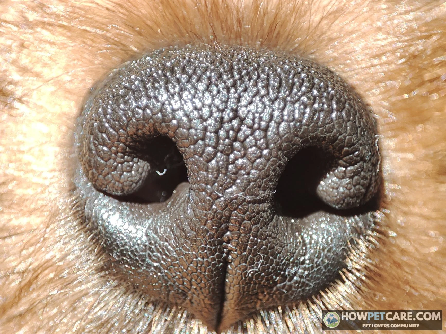
Do not apply human sunscreen to your dog’s nose without veterinary advice. Zinc oxide can be harmful if licked or swallowed, causing gastrointestinal irritation and, with substantial ingestion, potentially serious blood problems. Avoid products containing PABA as well. Ask your vet for a dog-labeled product or one they specifically approve, and how to prevent licking. If your dog eats sunscreen, contact your veterinarian or an animal poison-control service promptly.
Do not pick crusts or start medicated shampoos, ointments, or soaks without checking with the vet. A protective cone may help prevent scratching or face-rubbing if your vet recommends one.
Tracking lesions between vet visits
A brief weekly log can make a recheck more useful:
- Photograph the nose, ears, around the eyes, feet, and other affected areas in similar lighting.
- Note new pustules, spreading crusts, cracks, bleeding, or changes in walking.
- Record medication changes or missed doses, along with appetite, energy, drinking, and urination.
- Note vomiting, diarrhea, unusual odor or discharge, and possible urinary changes.
Call your vet within a day or two about new lesions in typical PF locations, spreading crusts, cracked pads, or signs returning during a taper. Call sooner if your dog seems unwell. Do not wait for the next routine visit or independently increase medication.
When a flare becomes an emergency
A few new crusts in an otherwise comfortable dog are not necessarily an emergency. Seek urgent veterinary advice or emergency care for:
- Collapse, severe weakness, or profound lethargy, especially with fever.
- Repeated vomiting or diarrhea, particularly if there is blood or black, tarry stool.
- Refusal to eat accompanied by illness, significant pain, or vomiting.
- Rapidly spreading, oozing, or very painful lesions, especially if your dog seems unwell.
- Extreme thirst and urination accompanied by weakness or other signs of illness.
Keep bedding and living areas clean, and report suspected skin or urinary infections. PF itself does not require isolating your dog, but follow your vet’s instructions if a contagious infection is suspected or if your immunosuppressed dog needs special precautions.
Your Next Steps: A Decision Guide for Owners Facing a PF Diagnosis
If your dog has suspicious crusts, book a veterinary visit, take dated photographs, and bring a list of medications and parasite preventives. Ask whether cytology, tests for infection, and biopsy are appropriate before treatment begins.
If PF has been diagnosed, use your next appointment to agree on a written plan:
- Confirm the diagnosis: What did the biopsy show, and were infections or other mimics checked?
- Understand treatment: Which medication is intended to control the flare, and is a steroid-sparing drug appropriate?
- Plan monitoring: When are the next examination, blood tests, and urine tests due? Which side effects require an immediate call?
- Discuss the budget: What is the estimated cost for the first three months, and what might a stable year of refills and monitoring cost?
- Set a referral point: Ask when a veterinary dermatologist should become involved if improvement stalls, side effects become severe, or relapses recur.
Also discuss routine preventive care. Vaccines may be less effective during immunosuppression, and timing depends on the vaccine, medication, local requirements, and your dog’s risk. Ask your vet about core and non-core vaccines and whether antibody titer testing is useful; do not cancel or schedule boosters without that discussion.
No diet or supplement has been proven to treat PF. Feed a complete diet suited to your dog, and check with your vet before adding supplements, including omega-3 products or herbs marketed to stimulate immunity. Your photographs, medication log, and observations about comfort and daily activities can help the veterinary team balance disease control against treatment side effects.
| Variant Name | Typical Lesion Locations | Lesion Type | Relative Frequency and Severity | Diagnostic Confirmation Method |
|---|---|---|---|---|
| Foliaceus | Face, ears, feet; sometimes widespread skin | Fragile pustules followed by crusts and erosions | Most common canine pemphigus form; severity varies | Examination, cytology, and biopsy |
| Erythematosus | Mainly face and nose | Localized crusting and sometimes pigment loss | Often milder and more localized | Biopsy and exclusion of similar diseases |
| Vulgaris | Mouth, lips, other mucocutaneous junctions, and sometimes skin | Deeper erosions or ulcers | Rare and often more severe; mouth pain can be significant | Biopsy showing a deeper pattern of skin-cell separation |
| Vegetans | Skin folds and other affected skin | Raised, wart-like lesions that may erode | Rare | Biopsy interpreted with the clinical appearance |
1What cellular structures are disrupted by autoantibodies in canine pemphigus foliaceus?
Autoantibodies attack desmosomes, causing skin cells to separate in a process known as acantholysis.
2Which diagnostic test is considered the key confirmatory step for pemphigus foliaceus when interpreted alongside history and exam findings?
Histopathology of tissue samples examined by a pathologist provides the key confirmatory test when interpreted alongside clinical findings.
3Where do early skin lesions of pemphigus foliaceus most commonly manifest on a dog?
Pemphigus foliaceus typically initiates on facial locations such as the nasal bridge, periocular skin, and pinnae before potentially spreading elsewhere.
4Why do dog owners more frequently notice yellow-tan crusts rather than intact pustules during a pemphigus foliaceus flare?
The separation of skin cells creates delicate blisters that easily burst, leaving behind characteristic yellow-tan crusts.
5How does pemphigus vulgaris primarily differ from pemphigus foliaceus in clinical presentation?
Pemphigus vulgaris targets deeper skin layers and mucocutaneous sites, often presenting with painful oral ulcerations.
6Why is human sunscreen containing zinc oxide unsafe for use on dogs with facial pemphigus foliaceus?
Dogs frequently lick topical substances, and swallowed zinc oxide poses toxic risks to the digestive tract and red blood cells.
7Which characteristic microscopic finding on skin cytology strongly supports a clinical suspicion of pemphigus foliaceus?
The presence of rounded, detached skin cells resulting from cell-to-cell separation supports a diagnosis of pemphigus foliaceus.
8Why must pet owners avoid abruptly stopping a dog's prescribed systemic steroid treatment?
Prolonged corticosteroid therapy suppresses normal adrenal gland function, making sudden withdrawal dangerous to bodily hormone regulation and skin stability.
9What is the main objective of adding a steroid-sparing immunosuppressant (such as azathioprine) to a dog's treatment plan?
Combining medications allows clinicians to achieve immune control while minimizing high-dose steroid complications over time.
10Based on veterinary outcome studies cited, what is the approximate complete remission rate reported for dogs treated for pemphigus foliaceus?
Published studies indicate complete clinical remission in roughly 52% of cases, though outcomes vary across individual dogs and study definitions.
FAQ
Can my dog still be vaccinated while on immunosuppressive medication?
It depends on the dog and the dose. Vaccines may be less effective during immunosuppression, and some vets limit vaccines in dogs with autoimmune disease. Discuss timing, core vs. non-core vaccines and titer testing with your vet or dermatologist before any booster.
Are there diet changes or supplements that help pemphigus foliaceus?
No diet or supplement has been proven to treat PF. Omega-3 fatty acids are sometimes used to support skin health, but evidence is limited. Check with your vet before adding any supplement, especially immune-stimulating herbs, which could interfere with treatment.
Does pemphigus foliaceus hurt or itch?
Itch varies, and some dogs have little at first. Crusted or cracked footpads and eroded skin can be painful, and dogs with widespread disease may feel unwell. Tell your vet if your dog seems sore, limps or stops eating.
Can pemphigus foliaceus turn into pemphigus vulgaris?
They are distinct diseases, and PF is not generally described as progressing into vulgaris. Vulgaris involves deeper blistering and mouth or mucous membrane lesions. A biopsy distinguishes them, so new mouth sores or ulcers should be reported to your vet.
Is pemphigus foliaceus painful enough to consider euthanasia?
Most dogs do not need euthanasia, but about 13% were euthanized in the cited outcome data, often because of poor response, side effects or cost. If your dog’s quality of life is falling, discuss treatment changes or a dermatology referral first.