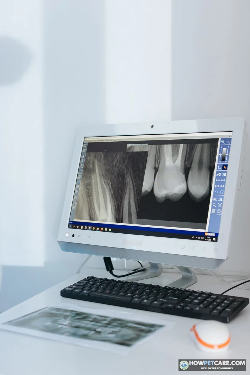

A cat X-ray—also called a veterinary radiograph—is a painless, noninvasive image showing bones, chest, organ outlines, gas, and many swallowed objects or stones. Most calm cats need no sedation, typical US general-practice costs run about $100–$300, and results are often discussed the same day.
A cat X-ray—also called a veterinary radiograph—is a quick, noninvasive image that helps a veterinarian look for broken bones, swallowed objects, bladder stones, and changes in the chest or abdomen. The exposure itself is painless, although positioning can be uncomfortable for an injured cat; many cats can be X-rayed without sedation.
What happens next depends on the vet’s goal. Ask for an estimate that covers the whole study, find out whether your cat may need sedation, and follow the clinic’s own preparation instructions. If you searched for “cat in x-ray” because you already have an image in hand, the five-step method below can help you follow your vet’s explanation—not diagnose the image yourself.
What a Cat X-Ray Shows: Chest, Abdomen, Skeleton and Teeth
What a cat X-ray shows by body region
An X-ray turns structures that absorb different amounts of radiation into a two-dimensional picture. Bones and many mineralized objects stand out; air-filled spaces look dark. Soft-tissue organs appear as outlines, but their internal detail is limited.
- Chest: A cat chest X-ray can show the size and shape of the heart, lung patterns, fluid around the lungs, masses, and some airway changes. A vet may recommend it for coughing, suspected heart or lung disease, or breathing problems once the cat is stable enough to be positioned.
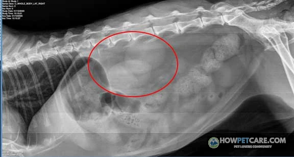
- Abdomen: A cat abdominal X-ray can help assess the stomach, intestines, bladder, kidneys, and organ outlines. It may reveal gas patterns linked to a bowel obstruction, certain bladder stones, or a swallowed object.
- Skeleton: Images of a limb, pelvis, or spine can help identify fractures, dislocations, arthritis, and other bone or joint changes.
- Teeth: Dental radiographs show tooth roots and surrounding bone beneath the gumline. They are commonly taken with a dental X-ray unit while the cat is under general anesthesia.
Signs your cat may need an X-ray
Your vet decides whether imaging is useful after considering the exam and history. Repeated vomiting, not eating, limping, coughing, a fall, or a possible swallowed object are all reasons to contact a vet rather than rely on an image online.
Get emergency veterinary care for open-mouth or labored breathing, major trauma, or repeated attempts to urinate with little or no urine. A suspected swallowed object with repeated vomiting also needs urgent assessment. Call ahead if you can, but don’t delay care to arrange an X-ray.
A mild limp or occasional cough in an otherwise comfortable cat may be suitable for a prompt regular appointment; ask your clinic how soon your own cat should be seen. Urinary straining can be mistaken for constipation, so contact a vet immediately if you’re unsure whether urine is being produced.
What an X-ray can’t tell you
A radiograph narrows possibilities; it doesn’t give a complete picture of every tissue. It may miss string, fabric, certain stones, or subtle changes inside an organ. Ultrasound, bloodwork, CT, or another test may be needed. An X-ray can show a suspicious mass, but it can’t by itself establish that the mass is cancer.
Are Cat X-Rays Safe? Radiation, Lead Aprons and Staying in the Room
How much radiation is involved
Diagnostic X-rays use ionizing radiation in a brief exposure. The actual dose depends on the area examined, the equipment, and the number of images; no single dose fits every cat X-ray. For a medically justified study, the diagnostic benefit generally outweighs the small radiation risk.
Staff limit unnecessary exposure by selecting appropriate settings, narrowing the beam to the area of interest, and avoiding needless repeats. Digital radiography makes images available quickly and easy to share, although the dose still depends on the technique used. No fixed number of lifetime X-rays is right for every cat: repeat studies should have a clinical purpose, such as checking fracture healing.
Will the clinic let you stay in the room?
Many clinics ask owners to wait outside during the exposure. Policies depend on local radiation-safety rules, the facility, and whether an owner’s presence is necessary. Being asked to leave doesn’t mean something has gone wrong.
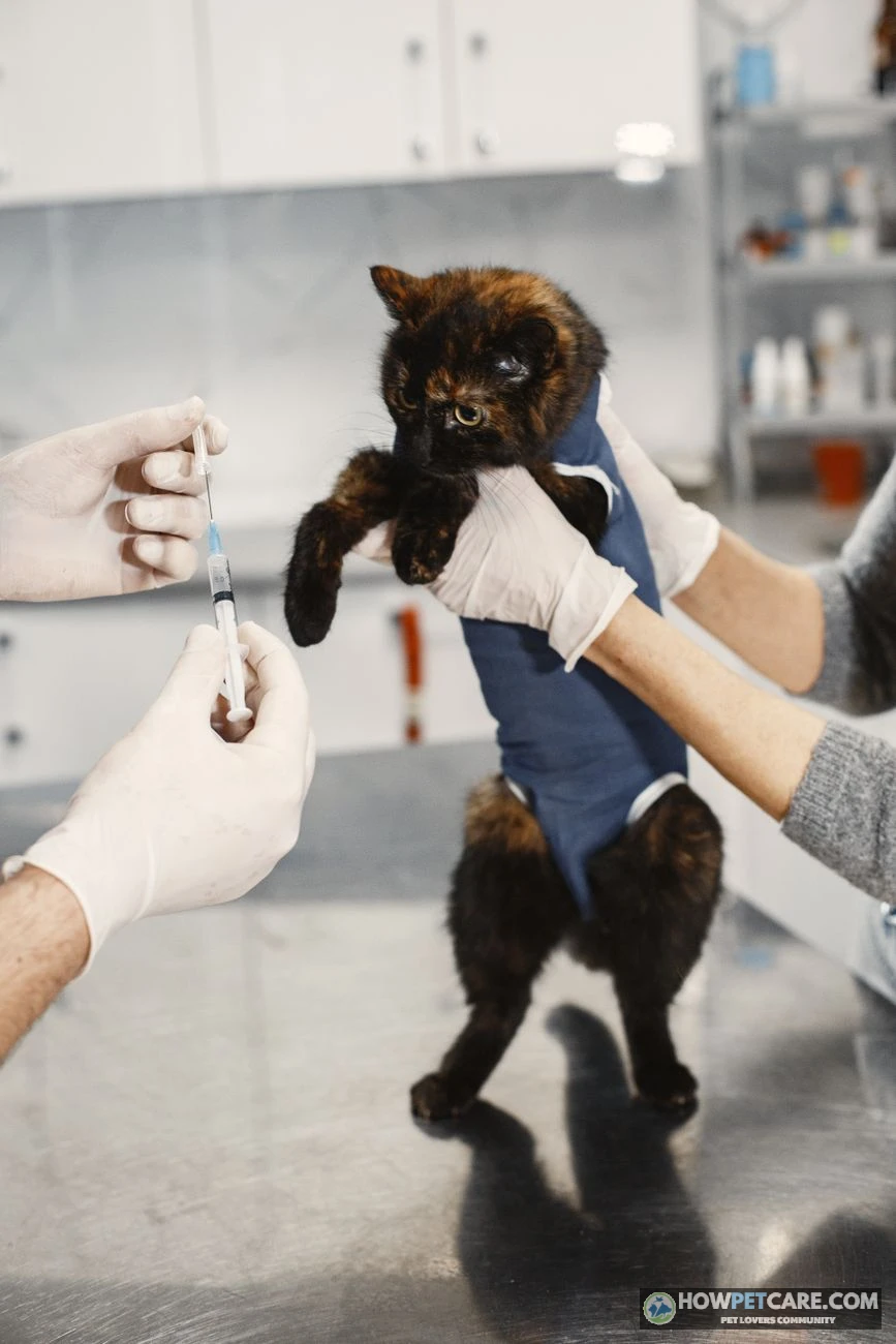
Staff may use foam wedges, troughs, sandbags, or other positioning aids to reduce hands-on restraint. Sedation is another option when it makes positioning safer or less stressful.
Lead aprons and thyroid shields for owners
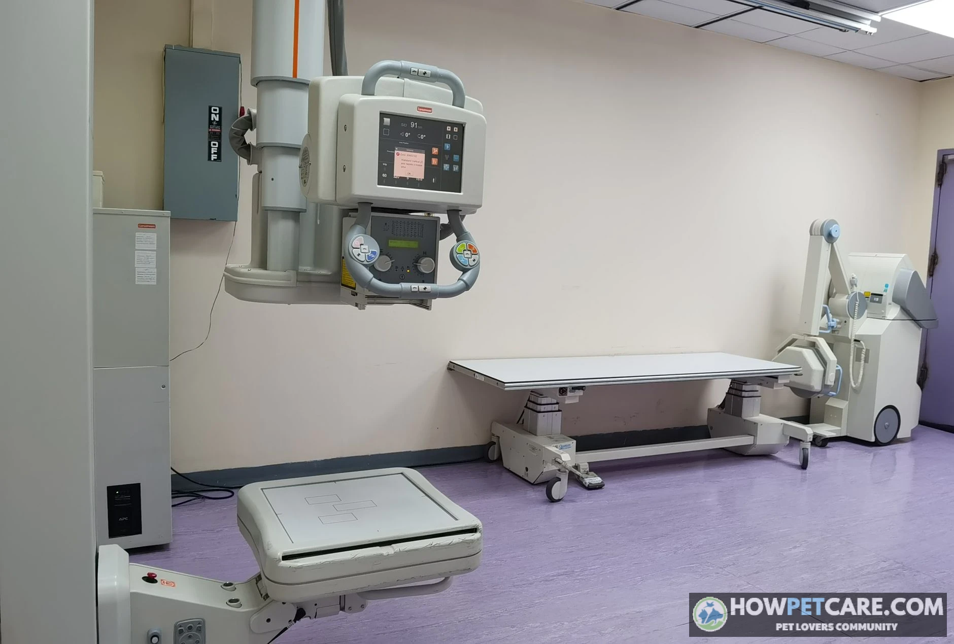
If a clinic lets you help in the room, follow its instructions exactly. Protective equipment may include a lead apron and thyroid shield; gloves and other protection may also be required. Veterinary personnel who restrain animals use protective equipment and exposure monitoring.
Children and pregnant people are generally asked not to assist with restraint. Tell the clinic in advance if that applies to you, and ask about its policy before the appointment.

 You might be wonderingCat yearsCat Years to Human Years: Conversion Chart & Age GuideRead the answer
Does Your Cat Need Sedation for an X-Ray? A Decision Guide
Sedation is about getting a still, correctly positioned image while managing stress and pain—not about the X-ray beam. A calm cat may have chest or abdominal images taken awake; another cat having the same study may need medication. Pain, movement, the area being imaged, and the cat’s health all shape the decision.
Sedation vs. anesthesia in plain terms
Sedation reduces awareness and anxiety to a degree that depends on the drugs and dose. General anesthesia produces a deeper, controlled loss of consciousness and requires appropriate monitoring and airway management. A sedated cat can still need separate pain relief; sedation doesn’t replace an analgesic.
When sedation is usually needed and when it isn’t
| Scenario | Sedation usually needed? | Why | What to ask your vet |
|---|---|---|---|
| Calm adult cat; routine chest or abdominal study | Often no | A relaxed cat may tolerate gentle positioning. | “Can we try awake if that is safe and comfortable?” |
| Suspected fracture or painful limb | Often helpful | Positioning can hurt, and movement can blur the image. | “What pain relief will my cat receive?” |
| Very anxious or struggling cat | Often helpful | Medication can reduce distress and make positioning safer. | “Which approach would be least stressful?” |
| Dental radiographs | General anesthesia is normally used | Sensors must be placed accurately inside the mouth. | “Is anesthesia included in the dental estimate?” |
| Skull or spine study | Sometimes sedation or anesthesia | Precise positioning may be difficult while awake. | “How much positioning will be needed?” |
| Breathing difficulty | Case by case; stabilize first | Handling and medication both require careful assessment. | “How will you stabilize my cat before imaging?” |
| Late-pregnancy kitten count | Often no | Many cats tolerate a brief abdominal study awake. | “Can this be done comfortably without sedation?” |
Questions to ask before you agree
Sedation and anesthesia carry a low but real risk. Cats with heart, lung, liver, or kidney disease may need a modified plan or extra assessment; the vet may recommend bloodwork as part of that assessment. Bloodwork doesn’t eliminate risk or reveal every heart or breathing problem.
Ask which drugs are planned, how your cat will be monitored, whether bloodwork is recommended, and what the extra charges cover. Tell the team about previous reactions to medication and any known medical conditions.
How Much Does a Cat X-Ray Cost? Price Ranges by Region, Sedation and Clinic Type
Typical US price ranges
For budgeting, the following are illustrative, unverified planning ranges, not a current survey or a quote from a specific clinic. Published fee schedules were not available in the supplied evidence, so these figures should not be treated as verified 2026 market prices. A local written estimate is essential, particularly for emergency care.
| Body region or add-on | Views typically taken | General practice range (USD) | Emergency or specialty range (USD) | What affects the price |
|---|---|---|---|---|
| Chest or abdomen | 2–3 | $100–$300 | $250–$600 | Views, facility, interpretation, and timing |
| Limb or single joint | Often 2 | $80–$250 | $200–$450 | Positioning, injury, and sedation needs |
| Multiple regions or additional series | 3 or more | $200–$450 | $400–$800 | Number of areas and complexity |
| Full-mouth dental radiographs | Multiple dental views | $150–$400 | $300–$700 | May be bundled into a dental procedure; anesthesia is additional unless specified |
| Sedation or anesthesia | Not applicable | $50–$250 | $150–$400 | Drug protocol and monitoring; general anesthesia can cost more |
| Pre-sedation bloodwork | Not applicable | $80–$150 | $100–$250 | Tests included and clinic pricing |
| Specialist radiologist review | Not applicable | $80–$200 | Ask the clinic | May be billed separately or included |
These rows aren’t interchangeable packages, and the ranges shouldn’t be added automatically: clinics bundle services differently. A dental procedure involving anesthesia, monitoring, cleaning, and treatment can cost substantially more than the radiograph line alone.
What adds to the bill
The final bill may include an exam, emergency or after-hours fee, pain medication, sedation, anesthesia and monitoring, bloodwork, additional views, and a radiologist’s interpretation. An ultrasound, contrast study, hospitalization, or treatment for a diagnosed problem is generally separate unless the estimate says otherwise.
Location, staffing, equipment, and clinic policies all influence the total. An emergency or specialty hospital may charge more than a general practice, but neither an urban address nor an independent ownership model guarantees a particular price.
Ways to keep the total manageable
Ask: “What is the estimated total for the exam and the views you expect, including any likely sedation and monitoring?” Request a low-to-high written estimate and ask what circumstances would change it. If cost is a constraint, tell the vet before testing so you can discuss priorities without delaying urgent care.
Ask whether a radiologist’s review is included and whether the clinic offers payment options; availability varies. Pet insurance may reimburse diagnostic imaging for a covered condition, subject to exclusions, deductibles, and policy terms. Pre-existing conditions are commonly excluded.
Before and After: Cat X-Ray Prep Checklist and Sedation Recovery Timeline
Prep checklist: fasting, what to bring, and carrier tips
- ☐ Confirm food instructions. Fasting may be requested when sedation or anesthesia is planned, but an awake X-ray doesn’t automatically require it. Don’t impose your own fasting schedule.
- ☐ Ask about water and medication. Don’t withhold either unless the clinic directs you to. Kittens and cats with diabetes need individual instructions.
- ☐ Gather records. Bring a medication and supplement list, relevant medical history, and previous images or reports if you have them.
- ☐ Write down symptoms. Note when they began, what has changed, and whether your cat is eating, vomiting, or urinating.
- ☐ For a possible swallowed object, bring details. A photo or packaging can help identify what may have been eaten and when.
- ☐ Use a secure carrier. A familiar blanket or towel can make the trip less stressful. If you use a feline pheromone spray, apply it to the bedding as directed on the product—not directly to the cat.
- ☐ Ask about the estimate and plan. Confirm expected sedation, who will read the images, and whether you can receive a digital copy.
For an emergency, call on the way if you can; don’t delay the trip to collect records or complete a fasting period.
What happens during the appointment
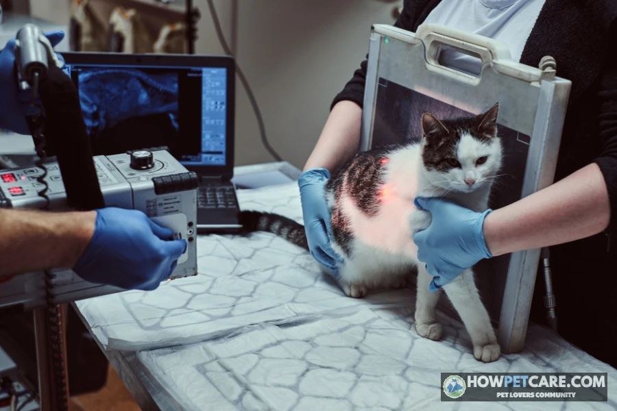
The team checks your cat’s history and condition, then positions the relevant body part for the prescribed views. The exposure itself is brief; setup and positioning take longer. A straightforward awake study may be completed during the visit, while medication, emergency stabilization, or a specialist review adds time.
Your veterinarian may discuss an initial interpretation the same day. A radiologist’s report can take longer, depending on the service and urgency. Ask when to expect results and how the clinic will contact you.
Home care and warning signs after sedation
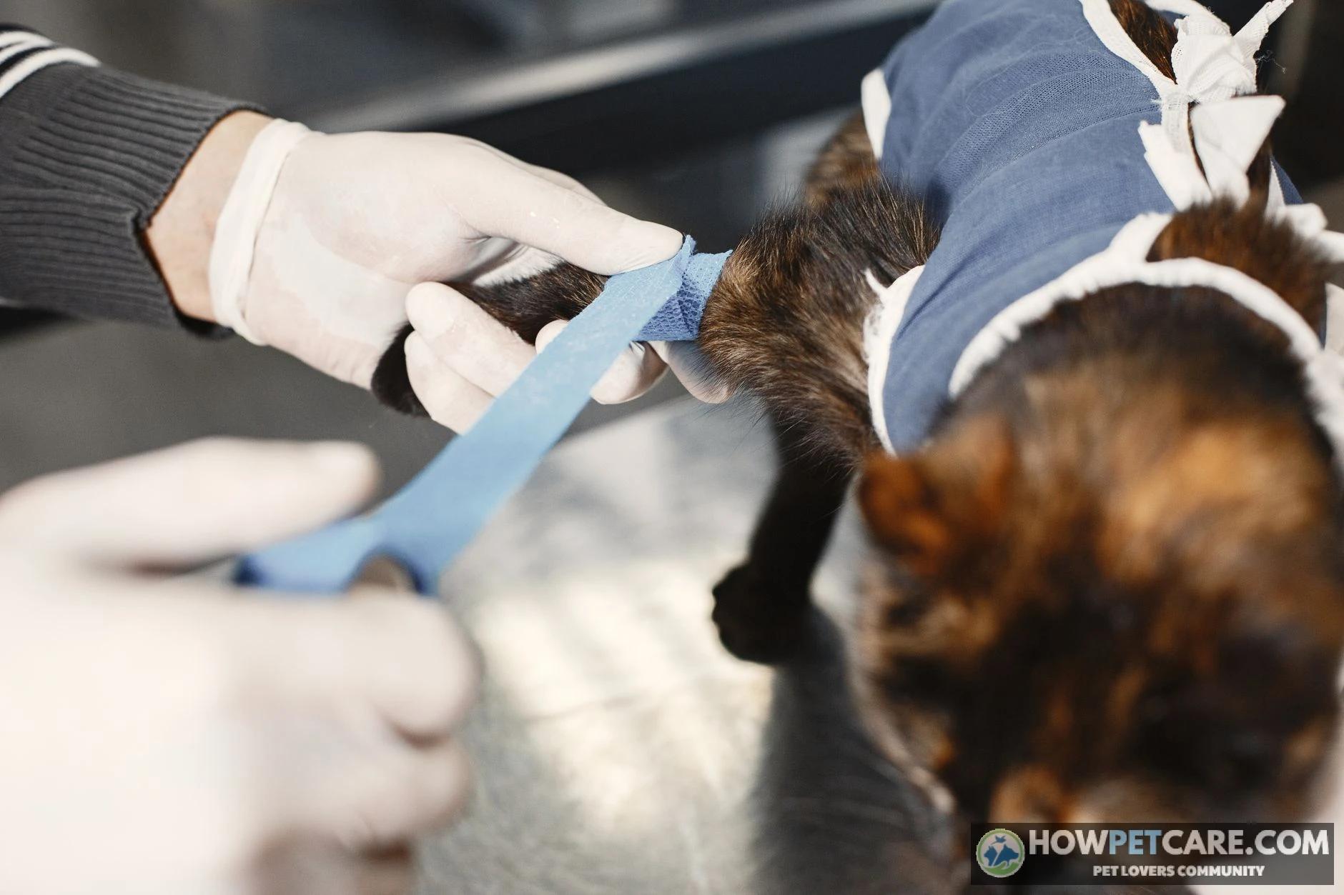
After an awake X-ray, most cats can return to their usual routine and eat normally unless the vet gives different instructions. After sedation or anesthesia, follow the discharge sheet for food, water, medication, and activity.
During the first several hours: Sleepiness and some wobbliness can occur. Keep your cat indoors in a quiet, warm room with a low bed and easy access to a litter box. Prevent jumping or stair use while coordination is impaired. Offer food only when and in the amount your clinic recommends; a small meal may be advised.
Later that day and overnight: Your cat should become progressively more alert. Recovery varies with the medication and the individual cat, so call the clinic if grogginess isn’t improving as expected or you’re worried.

Contact the clinic immediately or go to an emergency vet for trouble breathing, inability to rouse your cat, pale gums, repeated vomiting, extreme weakness, or worsening rather than improving responsiveness. Urinary straining with little or no urine is also an emergency.
If your cat hasn’t urinated after returning home, call the clinic for advice, especially if the cat is uncomfortable or making repeated attempts. Before leaving the appointment, save the clinic’s after-hours number.
How to Read a Cat X-Ray: A Five-Step Method With Worked Abnormal Examples
A cat X-ray image is a flattened view of overlapping structures. Use this method to organize questions for your veterinarian, not to decide that an image is normal or to make a diagnosis. The vet or a veterinary radiologist interprets the study alongside the exam, history, and other tests.
Step 1 to 5: the beginner reading checklist
- Orient the image. Find the view label and the right or left marker. A lateral view is taken from the side. A dorsoventral or DV view has the cat positioned belly-down; a ventrodorsal or VD view has the cat belly-up. Ask which end is the head if it isn’t obvious.
- Learn the density code. Air generally appears black, fat dark gray, soft tissue and fluid gray, and bone white. Metal is usually bright white; mineralized material such as some stones can also look bright. Display settings and overlap affect appearances, so color alone doesn’t identify a structure.
- Scan the outline and chest. Trace the body edge, ribs, and diaphragm—the boundary between chest and abdomen. Air-filled lungs generally look darker than the heart and surrounding soft tissue, with visible vessel markings. Ask the vet to point out the heart, windpipe, and any area that looks unusually opaque.
- Use reference points, not guesses. Compare corresponding sides and follow the spine and ribs. Vets may use a vertebral heart score, comparing heart dimensions with vertebral lengths, as one aid when evaluating heart size. Positioning and individual variation matter; the measurement isn’t a diagnosis, and an apparently normal heart silhouette doesn’t rule out heart disease.
- Move through the abdomen and skeleton. Ask the vet to identify the stomach, intestines, colon, bladder, kidneys where visible, and bones. Notice whether bowel loops differ markedly in diameter or contain gas, but don’t assume that a dark pocket or bright spot is abnormal without the vet’s interpretation.
Why the vet takes lateral and dorsoventral views
One image compresses a three-dimensional body into a flat picture. Structures can overlap and disguise one another—a normal effect called summation. For example, the kidneys can overlap on a side view.
Views from different directions help the vet locate a finding and distinguish overlap from a true abnormality. A chest study often includes both side views and a DV or VD view, because changing position can reveal details that one image hides. Ask which views were taken and what each adds.
What a swallowed object or bowel obstruction looks like
Imagine a side view of the abdomen with several dark, gas-filled intestinal loops. If some loops are enlarged and others are smaller, the pattern may prompt the vet to investigate an obstruction. A metal object may be conspicuously bright; string, fabric, and some plastic can be difficult or impossible to see directly.
That pattern isn’t proof of a blockage, and an image without an obvious object doesn’t rule one out. Depending on the case, the vet may recommend ultrasound, repeat images, or a contrast study. A barium contrast study uses contrast material and a series of radiographs to help investigate the digestive tract when appropriate.
What bladder stones and fractures look like
Bladder stones: Some appear as bright, rounded mineral shapes in the region of the bladder. Others aren’t reliably visible on plain X-rays. A bright spot can also represent something outside the bladder or an overlapping structure, so its location and significance need professional assessment.
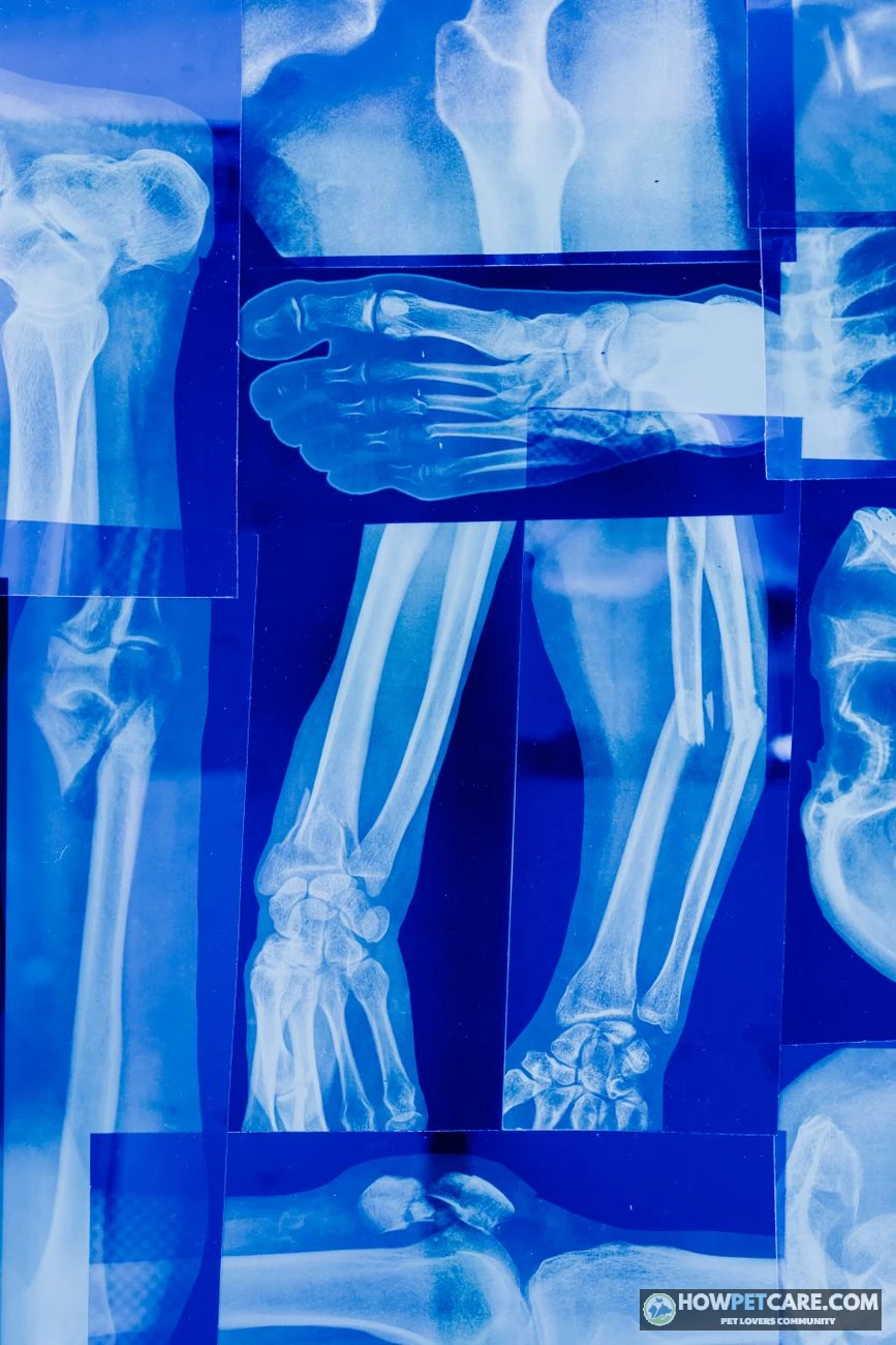
Fracture: A break may appear as a dark line crossing a white bone, an interruption in the bone’s smooth outline, or a segment that is angled or displaced. Some fractures are subtle and require several views.
Chest change: Compared with darker air-filled lung, fluid or other abnormal material can make part of the chest appear more opaque. A heart silhouette that looks enlarged can also prompt further testing, but an X-ray alone doesn’t establish the cause. After trauma, a diaphragmatic hernia can allow abdominal organs into the chest and may be visible on a radiograph.
Request a digital copy and ask your vet to point out one normal structure and the finding of concern on each view. If the finding remains unclear, ask whether a radiologist’s review would help.
Pregnant Cat X-Ray: Counting Kittens, Timing and Birth Planning
When can an X-ray count kittens?
Yes. A pregnant cat X-ray can estimate the number of kittens once their skeletons have mineralized enough to show on the image. Vets commonly plan a counting radiograph after roughly day 45 of pregnancy; a later study may make the skeletons easier to distinguish. If mating dates are uncertain, tell the vet.
The veterinarian counts visible skulls and spines, but overlapping kittens can make an exact count difficult. Treat the result as an estimate, not a guarantee that every kitten has been counted.

X-ray vs. ultrasound for pregnancy
Ultrasound can detect pregnancy earlier and assess fetal heartbeats, while a late-pregnancy radiograph is useful for counting mineralized skeletons. The tests answer different questions, and a vet may recommend both. A single late-pregnancy radiograph may be justified when its expected benefit outweighs its small radiation risk; discuss timing and necessity with your veterinarian.
Many pregnant cats can be positioned awake, although the decision depends on temperament and health. Ask the clinic what to expect before the visit.
Using the count to plan delivery
An estimated kitten count gives you and your vet a reference during birth. It can help guide decisions if labor stops and a kitten may remain, or if delivery is difficult and a cesarean section is being considered. It doesn’t predict exactly how labor will proceed.
Ask your vet in advance which signs require an immediate call. Strong, unproductive straining, a kitten visibly stuck, a distressed mother, or concern that labor isn’t progressing warrants urgent veterinary advice.
Cat X-Ray vs. Ultrasound vs. CT: Which Imaging Does Your Cat Need?
Imaging comparison at a glance
| Imaging type | Shows best | Main limits | Sedation or anesthesia | Best-fit scenarios |
|---|---|---|---|---|
| X-ray | Bones, chest, organ outlines, gas, and many mineralized objects | Structures overlap; limited internal soft-tissue detail; some objects and stones are missed | Often none; sedation depends on the cat and views | Limping, chest assessment, suspected swallowed object, late kitten count |
| Ultrasound | Internal organ structure, fluid, intestinal walls, and fetal heartbeats | Air and bone limit the view; findings depend on the examination and operator | Often awake; some cats need sedation | Unclear abdominal findings, bladder or kidney assessment, pregnancy viability, heart ultrasound |
| CT | Cross-sectional detail of complex anatomy, including skull, nasal passages, spine, and masses | Greater expense, limited availability, and radiation exposure | Usually general anesthesia | Complex fractures, surgical planning, selected masses and nasal disease |
X-rays are generally less expensive than ultrasound, CT, or MRI, although the total for any individual workup depends on the clinic and associated care. MRI is another specialist option, particularly when detailed assessment of certain soft tissues or the nervous system is needed.
Which scan for which symptom
- Vomiting or a possible swallowed object: An abdominal X-ray may be a useful first test. Ultrasound can add information when the radiograph is unclear or soft-tissue detail matters.
- Limping or suspected fracture: X-rays are commonly the starting point. Complex injuries may need CT for planning.
- Breathing difficulty: Stabilization comes first. A vet may then recommend chest radiographs and, depending on the suspected cause, an ultrasound of the heart or chest.
- Urinary straining: This can be an emergency. Imaging may help identify stones or assess the bladder and kidneys, but the immediate priority is assessment and treatment.
The order isn’t fixed: the vet may choose ultrasound first when it’s better suited to the question. Ask, “What will this test show that the other test might not?” Referral may help with unclear findings, suspected masses, surgical planning, or access to advanced imaging.
Digital X-ray and AI-assisted reading
Digital radiography makes images easy to store and send for a remote specialist opinion, often called teleradiology. Some practices may also use AI-assisted tools to flag or support review of findings. These tools don’t replace the veterinarian’s clinical judgment or a radiologist’s interpretation. Ask who will read your cat’s images and whether a specialist review is recommended.
Your Next Steps: A Decision Checklist for Your Cat’s X-Ray
- Decide on urgency. Seek emergency care for labored or open-mouth breathing, inability to urinate, major trauma, or a suspected swallowed object with repeated vomiting. For less urgent signs, call your regular vet promptly for guidance.
- Prepare for the visit. Ask about the written estimate, fasting, medication, sedation, room policy, and who will interpret the study. Gather previous records and details of any possible swallowed item.
- At the appointment, ask for context. Which views were taken? What does each show? Does the finding explain the symptoms, and would bloodwork, ultrasound, or a radiologist’s review help?
- Before going home, get the plan. Request your discharge instructions, an after-hours contact, and a copy of the digital images. If your cat was sedated, confirm feeding and activity instructions and the signs that should prompt a call.
- Follow up if questions remain. Use the five-step viewing method to follow—not replace—the veterinary explanation. If the diagnosis is uncertain or treatment decisions are difficult, ask about a second opinion or specialist referral.
 You might be wonderingAnemia in catsAnemia in Cats: Symptoms, Causes, Costs & When It’s an EmergencyRead the answer
FAQ
How long does a cat X-ray take?
The exposure itself lasts a fraction of a second. Positioning and setup usually make the whole procedure take a few minutes to a few tens of minutes. A sedated cat needs additional time for the drugs to work and to recover.
Can a cat X-ray show cancer?
An X-ray can show masses, enlarged organs, and bone changes that raise concern, especially in the chest. It cannot confirm cancer on its own, so vets usually follow up with ultrasound, a biopsy, or other tests.
Will I get a copy of my cat’s X-ray images?
Most clinics store digital radiographs and can email them or put them on a disc or USB drive on request. Ask at check-out, especially if you are seeing a specialist or planning a second opinion.
How often can a cat safely have X-rays?
There is no set number. A veterinarian orders repeat X-rays when the information will change treatment, such as monitoring a fracture heal or a chest condition. Each study uses a low dose, and vets avoid unnecessary repeats.
Does pet insurance cover cat X-rays?
Accident-and-illness plans commonly reimburse X-rays when they are used to diagnose a covered condition or injury, and usually exclude pre-existing conditions. Routine or wellness imaging depends on the plan, so check the policy and deductible before the visit.