
How urgent a swollen lymph node is depends on the cat, not just the lump:
- Go to a vet today for labored breathing, no eating or drinking for ~24 hours, collapse, or a hot, painful, fast-growing swelling.
- Book a routine visit within days if a node stays enlarged past a week, feels firm or fixed, or several nodes are enlarged.
- Monitor at home for 1–2 weeks only for a small, soft node after a vaccine or mild cold in an otherwise normal cat.
- Set a day-14 reminder — if the node isn’t clearly shrinking, book a vet visit; a needle sample is what identifies the cause.
Swollen Lymph Nodes in Cats: Emergency, Vet Visit, or Watch-and-Wait?
Most swollen lymph nodes in cats are not an emergency. A single soft, mildly enlarged node in a cat that is still eating, grooming, and using the litter box is usually left over from an infection or a recent vaccination. Reactive swelling like this commonly settles within two to four weeks once the trigger clears. Other signs need action today: labored breathing, a cat that has stopped eating, collapse, or swelling that appeared within hours and is hot and painful to the touch.
There’s an important limitation, though. A swollen lymph node — lymphadenopathy — is not a disease. It’s a symptom of an underlying condition such as infection, inflammation, or cancer, and you can’t tell which one you’re dealing with by feeling through the skin. A firm node can be an abscess. A painless one can still be cancer. In the first ten minutes, focus less on identifying the lump and more on deciding how quickly your cat needs help.
Run this checklist before you read further. It takes about a minute and helps you decide when to act.
Work down the list from top to bottom. The first box you tick sets your deadline.
GO TO A VET TODAY (emergency clinic or same-day appointment)
- Your cat is struggling to breathe, breathing with an open mouth, or breathing fast at rest
- Your cat has not eaten or drunk anything for roughly 24 hours
- Your cat has collapsed, is extremely lethargic, or is hiding and unresponsive
- The swelling appeared fast (hours to a day), is visibly growing, or is hot and painful to a light touch
- Your cat feels hot or seems feverish, or the swelling is draining pus or blood
BOOK A ROUTINE VISIT WITHIN THE NEXT FEW DAYS
- A node has been enlarged for more than about a week with no improvement
- The node is growing, feels firm, or does not move freely under the skin
- You can feel more than one enlarged node
- Alongside the node, your cat has weight loss, reduced appetite, vomiting or diarrhea, sneezing, or eye/nose discharge
REASONABLE TO MONITOR AT HOME (re-check in 1–2 weeks)
- A small, soft, mildly enlarged node appeared within a week or two of a vaccination
- Mild node swelling appeared during a cold/upper respiratory infection that is already improving
- Your cat is otherwise normal: eating, drinking, grooming, playing, and using the litter box normally
Monitoring rule: set a phone reminder for day 14. If the node is not clearly shrinking — or is bigger — book a routine visit. Confirm the timing with your own vet.
If you ticked nothing on the list, you are in the third tier and have room to observe — but observation has rules, covered below.
Red flags that mean go to a vet today
Breathing difficulty, a cat that has stopped eating and drinking, collapse, and swelling that is hot, painful, or visibly expanding within hours should not wait for a Tuesday appointment. Rapid, painful, hot swelling is a classic sign of infection or an abscess, and an abscess near the throat or jaw can compromise the airway. Fever along with a swollen node also calls for a same-day call. If you’re unsure which of two tiers applies, choose the faster one — a false alarm costs an exam fee, and waiting can cost much more.
When it’s reasonable to monitor for a short time
Monitoring makes sense only with a deadline. Re-check the node at 1–2 weeks and book a visit if it hasn’t clearly started shrinking. Reactive nodes commonly take two to four weeks to resolve after the cause improves, so a node that’s smaller at day 14 is on the right track even if it hasn’t vanished. If it’s the same size, bigger, or firmer at the two-week mark, book a routine vet visit. Vaccination is the most common benign trigger in this tier: a vaccine or allergic reaction can stimulate the immune system and produce temporary reactive swelling. These reactions are usually mild and short-lived.
Be honest about what "otherwise normal" means for your cat. Your cat should be eating its usual amount, drinking, grooming, moving and jumping without stiffness, interacting with you, and producing normal urine and stool. A cat that’s quiet but still eating and using the box isn’t quite "normal." That’s a gray zone, and a gray-zone cat with a growing lump should be seen rather than watched.
The sections ahead explain how to find each node, distinguish it from other lumps, understand possible causes, and prepare for the vet visit and its costs in 2026 dollars.
How to Find and Gently Check Your Cat’s Lymph Nodes at Home
You can reach five pairs of nodes on a cat, enough to gather useful information before a vet visit. You can’t diagnose the cause by touch, though. You can’t feel an internal node, or distinguish reactive tissue from lymphoma. Aim for a careful map and a dated record, not a verdict.
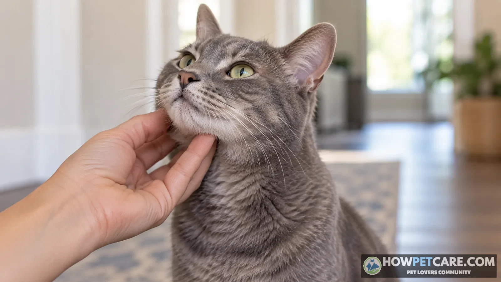
-
Set the room and the mood
Choose a quiet room, warm (not cold) hands, and keep treats within reach. Check your cat when it’s already relaxed, not after a chase.
-
Use the flats of two or three fingertips
Use feather-light pressure, as if finding a grain of rice under a sheet. Pressing hard tells you nothing and hurts.
-
Compare left to right, every time
Most cats have slight asymmetry, but a node that’s clearly bigger on one side is worth noting.
-
Stop the moment the cat objects
Stop if your cat tenses, growls, flinches, or pulls away. Pain on light touch is useful information — note it and stop.
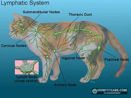
              [1] SUBMANDIBULAR — under each side of the jaw
     (head)        /
       |        /

  [2] PRESCAPULAR — just in front of each shoulder blade


  [3] AXILLARY — in the armpit (behind front leg)


  [4] INGUINAL — in the groin fold (in front of rear leg)


     (tail)  [5] POPLITEAL — behind each knee, on the back of the leg
Submandibular nodes (under the jaw)
Cup your fingers under the jawline near the angle of the jaw and slide gently from chin toward ear. A normal node here is pea-sized at most, and in many cats you can’t feel it as a distinct structure — you’ll feel soft tissue, not a marble. Owners often discover lumps here, but the area is also easy to misread: the salivary glands sit nearby, swollen glands near the jaw are easy to mistake for nodes, and swelling from bad teeth can show up here too. Note any lump larger than a pea, anything clearly bigger on one side, and any difference in how your cat tolerates touch on either side.
Prescapular nodes (in front of the shoulder)
Place your fingers in the soft hollow just ahead of the top of the shoulder blade and press lightly inward. A normal node is barely there. If you can roll a node between your fingers like a small grape, especially if it’s firm, measure and photograph it.
Popliteal nodes (behind the knee)
A lump behind the leg often sends owners searching for answers. With your cat standing or lying on its side, slide your fingers into the soft pocket directly behind the knee joint and pinch gently between finger and thumb. This is the easiest node to find in a healthy cat; a normal one feels like a small, soft pea. Have it checked if it’s noticeably bigger than pea-sized, firm, or enlarged on only one leg. This node drains the lower leg and often reacts to a wound or bite below the knee.
Axillary and inguinal nodes (armpit and groin)
These nodes are hard to evaluate. Lift the front leg slightly forward and feel high into the armpit for the axillary node; feel along the crease where the rear leg meets the belly for the inguinal node. In most cats, neither is detectable, and fat and skin folds can mimic lumps. If you find something obvious in either spot, record it and let the vet confirm what it is — don’t dig.
What "enlarged" feels like versus normal
If a lump is hot, very sore, soft and squishy, or has a scab or drainage point, stop handling it. Rupturing an abscess at home creates a mess and a bigger infection. Cover it if it’s draining, keep your cat calm, and call the vet.
Look for a node that’s noticeably larger than a pea or bean, different from the matching node on the other side, firm rather than soft, fixed rather than freely movable under the skin, or painful or warm. These are guides, not rules: a big soft node can be reactive, while a small firm one can be cancerous. After each check, record the location, side (left, right, or both), size compared with a familiar object (rice grain, pea, grape, walnut), whether it’s soft or firm, whether it moves or feels stuck, and the date. Take a phone photo with a coin beside the lump for scale. A dated photo is more useful than memory at the vet visit.
Also note whether you’ve found one enlarged node or several. A single enlarged node is localized and usually points to a problem in its drainage area, such as a bad tooth, an ear infection, or a bite on that leg. Several enlarged nodes in different regions are considered generalized and shift suspicion toward systemic infection or cancer. The pattern affects what your vet tests for, so be sure to mention it.
Is the Lump Under Your Cat’s Jaw or Neck Really a Lymph Node?
Often it isn’t. What an owner believes is a lymph node may be a salivary gland, a tooth-root abscess, or a harmless fatty lump. The difference affects both the urgency and the tests. Use the table to organize what you’ve noticed, then let the vet confirm it.
| Clue | Swollen lymph node | Salivary gland (mucocele) | Dental/tooth-root abscess | Lipoma (fatty lump) | Bite-wound abscess |
|---|---|---|---|---|---|
| Typical location | Under the jaw angle, in front of the shoulder, behind the knee | Under jaw or along the throat, often near the midline | On the face, cheek, or along the jawline itself | Anywhere on the body, often chest or belly | Neck, head, back, tail base — anywhere a bite landed |
| Texture | Firm to rubbery | Soft, squishy, fluid-filled | Firm swelling, may soften and rupture | Soft, doughy | Firm early, then fluctuant (fluid pocket) |
| Moves under skin? | Usually somewhat movable; fixed is a warning sign | Poorly defined, feels like a water balloon | No — anchored to jaw/bone | Yes, slides freely | Slightly, until it becomes tense |
| Painful? | Varies — often painless if cancerous, sore if infected | Usually not painful, but may bother eating | Yes — cat resents touch near the mouth | No | Yes — very sore, cat may hide |
| Speed of growth | Days to weeks | Slow (weeks) | Days | Slow (months) | Fast — 1–3 days |
| Telling extra signs | Several lumps, weight loss, appetite change | Drooling, trouble swallowing | Bad breath, drooling, dropping food, pawing at mouth | None — cat acts normal | Recent outdoor fight, fever, matted fur, oozing wound |
| Who gets it most | Any cat | Any cat | Cats with dental disease (common over age 3) | Middle-aged and older, often overweight cats | Outdoor and multi-cat households |
Any lump that is new, growing, or hard deserves a needle sample — no matter how confident you are about what it is. Owners are often surprised when apparent "lymph nodes" turn out to be salivary tissue, or "fatty lumps" turn out to be nodes.
Lymph node vs salivary gland swelling
A salivary mucocele is a collection of saliva that forms a soft, fluid-filled swelling under the jaw or along the throat. It tends to feel like a water balloon rather than a distinct oval, and it may come with drooling or difficulty swallowing. The submandibular node sits in the same region, which is why this area needs a professional examination and, often, a needle sample.
Lymph node vs dental abscess
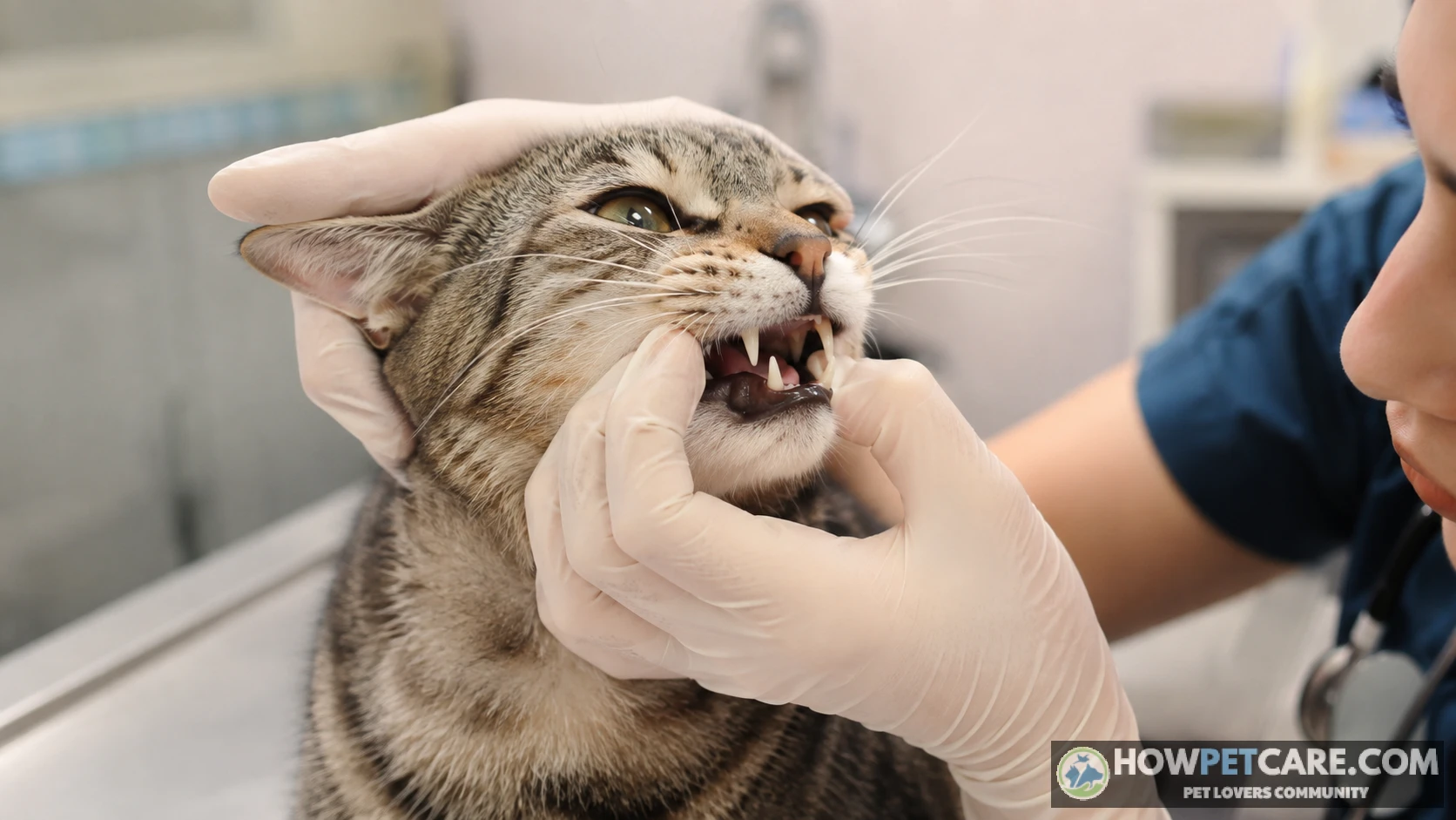
A tooth-root infection produces swelling anchored to the jawbone, usually along with bad breath, drooling, food-dropping, pawing at the mouth, or flinching when the muzzle is touched. The draining node under the jaw often enlarges in response to the dental infection, so both can be present at once. Dental disease is one of the most common underlying causes of a swollen submandibular node. Treating the node without treating the tooth fixes nothing.
Lymph node vs lipoma or cyst
A lipoma is soft, doughy, freely movable, and usually painless. It grows over months in a middle-aged or older cat. A cyst feels like a firm pea under the skin and may have a central pore. Both are far less concerning than a firm, fixed, fast-growing mass, but neither can be diagnosed by feel. Fine needle aspiration is quick, inexpensive relative to imaging, and settles the question in many cases.
What Causes Swollen Lymph Nodes in Cats, and How to Tell Reactive, Infectious, and Cancerous Apart
Lymph nodes are filters along the lymphatic system, the network that moves fluid and immune cells through the body. When a node detects inflammation, infection, or abnormal cells, it enlarges as immune cells pour in. That’s what happens in almost every swollen node you’ll feel on a cat.
Reactive and benign causes (including after vaccination)
Reactive hyperplasia is the technical name for a node responding to the immune system, and it’s the most common benign pattern. Triggers include recent vaccination or an allergic reaction, which can produce temporary reactive swelling, as well as a nearby skin wound, flea allergy, and dental inflammation. These nodes are usually soft, movable, and mildly enlarged. They shrink once the trigger resolves, commonly within two to four weeks.
Infectious causes
Infection is the most common cause of swollen lymph nodes in cats overall. Common sources include feline upper respiratory infection, an abscess from a bite wound, cat scratch disease (Bartonella), fungal disease such as histoplasmosis, and dental infection. FIV and FeLV matter too: the viruses can enlarge nodes directly, and they can weaken immune defenses enough to let other infections take hold. Tick exposure is another route. Cats without tick prevention carry a higher risk of lymph node enlargement from tick-borne diseases such as Lyme, Anaplasma, Ehrlichia, and Babesia.
Cancerous and immune-related causes
Lymphoma is the cancer most often linked to swollen nodes in cats, and the alimentary (intestinal) form is the most common presentation. Nodes can also enlarge when a cancer elsewhere spreads into them. Immune-mediated conditions can cause generalized node swelling without infection or cancer; sorting these out requires bloodwork and often a biopsy.
Node features can guide suspicion, but they never confirm a cause. A fine needle aspirate is the standard first diagnostic step for this reason. If your vet offers sampling at the first visit, take it — it can make the difference between treating a guess and treating a diagnosis.
| Feature | Reactive / benign | Infectious | Cancerous (e.g., lymphoma) |
|---|---|---|---|
| Typical size | Pea to small grape; mild enlargement | Pea to walnut; can be quite large | Pea to golf ball; keeps growing |
| Pain on touch | Usually not painful | Often painful, especially with abscess | Usually not painful — a painless growing node is the concerning pattern |
| Warmth | Normal temperature | Often warm or hot | Usually normal temperature |
| Mobility under skin | Soft, slides freely | Movable early; may stick if abscessing | Often firm, may feel fixed or "stuck" |
| Number affected | Usually one node near the trigger (e.g., jaw node after dental work) | One or a few near the infection site | Often several nodes at once, in different body areas |
| Duration | Days to ~2–4 weeks, then shrinks | Persists until infection is treated | Weeks to months, progressively enlarging |
| Company it keeps | Recent vaccine, skin wound, dental disease | Fever, discharge, lethargy, fight wounds, poor appetite | Weight loss, appetite change, possibly vomiting/diarrhea; cat may seem "off" without a clear infection |
| Common triggers | Vaccination, mild URI, flea allergy, dental inflammation | URI, bite abscess, Bartonella (cat scratch), fungal infection, FIV/FeLV-related infection | Feline lymphoma; cancer spread from another site; FeLV raises lymphoma risk |
Overlap warning: An infected node can be firm and fixed; early lymphoma can be small. This table can guide suspicion, but it cannot confirm or rule out cancer. The feature that most reliably points toward testing is duration plus trajectory: a node still enlarged after about two weeks, or one that’s still growing regardless of how it feels.
A swollen lymph node can be a sign of cancer — lymphoma is a recognized cause — but infection and reactive change are more common overall. There is no reliable published percentage showing how often a swollen node in a cat is cancer rather than infection, so a reassuring statistic offered at home would be a guess. Signs that raise concern include a firm, painless node that keeps growing, several enlarged nodes at once, and accompanying weight loss or appetite change. A painful, warm node that appeared quickly in a cat with a wound, a cold, or a recent vaccine is less concerning for cancer.
Keep this distinction in mind: a swollen lymph node in a cat is a symptom, not a diagnosis — and a needle sample, not the way it feels, is what identifies the cause.
How Vets Diagnose Swollen Lymph Nodes in Cats
Physical exam and history
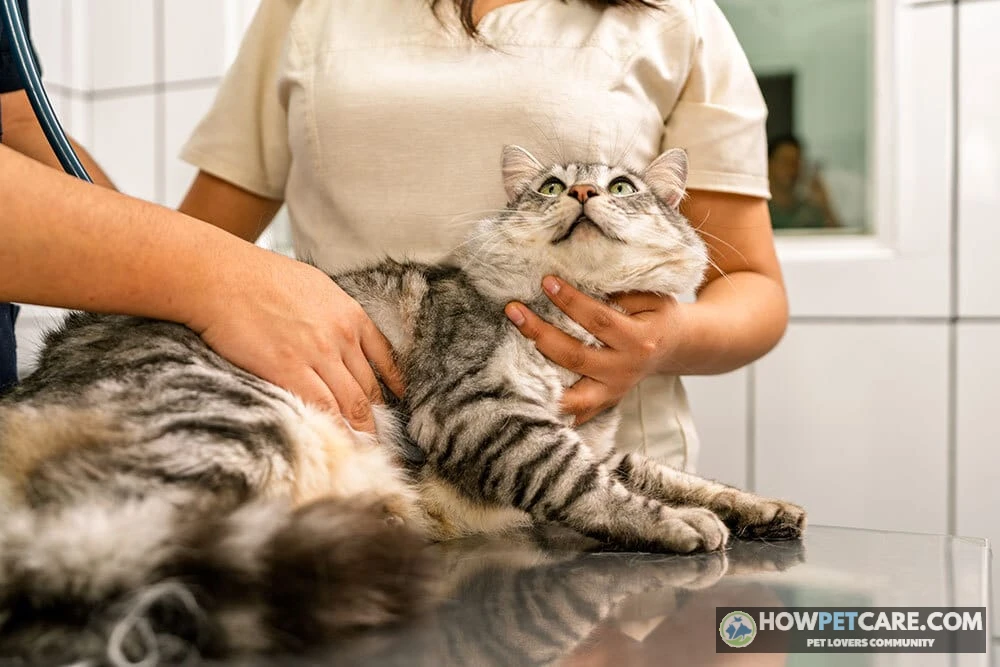
The exam starts with a full node map — all five peripheral pairs, not just the one you noticed — along with checks of the mouth, ears, skin, eyes, and body temperature. Your vet will ask when you first noticed the lump, whether it’s growing, whether your cat recently had a vaccine, fight, or illness, whether it goes outdoors, and whether it’s on flea and tick prevention. Your answers help guide the rest of the work-up.
Bloodwork and FeLV/FIV testing
A complete blood count and chemistry panel show whether there’s an active infection, inflammation, anemia, or organ problems; a urinalysis rounds out the picture. FeLV/FIV testing is standard rather than optional: viral status changes the differential list, the outlook, and how the cat should be managed around other cats.
Fine needle aspiration and cytology
FNA is the most common first diagnostic step for an enlarged node. A thin needle collects cells from the node, usually without sedation, and a pathologist or the clinic’s lab examines the slide. It often distinguishes reactive tissue from infection or suspected lymphoma. Cytology has limits, though: it reads individual cells, not tissue architecture, so some cases need a biopsy or immunophenotyping to confirm type and grade.
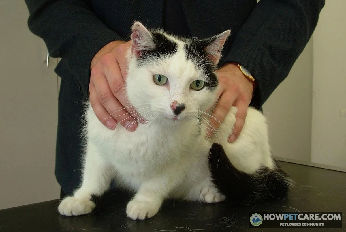
If the node is enlarged and your cat is healthy enough, "can we do an FNA today?" is a reasonable question. Waiting two weeks to see whether a node shrinks is fine for a soft node after a vaccine; it’s not the right plan for a firm node in a cat that’s losing weight.
Biopsy and imaging
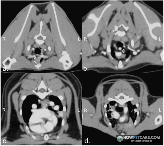
A biopsy involves surgical removal of a node or part of one, usually under sedation or anesthesia. It’s recommended when cytology is inconclusive or when lymphoma needs typing and grading, because it shows tissue architecture that cytology cannot. Imaging (chest X-rays and abdominal ultrasound) looks for enlarged internal nodes, organ involvement, and spread. It’s part of staging once lymphoma is suspected. Your vet may add tests such as Bartonella culture or PCR, or fungal testing, depending on your region, your cat’s history, and the first results.
Bring dated photos of the lump, a symptom log, the vaccine record, flea and tick prevention history, current medications, and information about whether your cat goes outdoors or has been in fights. Ask which nodes are enlarged, whether the vet can sample today, what the differential list is, which tests change treatment, and what each will cost.
What Swollen Lymph Node Testing Costs for Cats (US Ranges)
The figures below are approximate US ranges compiled from publicly posted clinic fee schedules and practice surveys, last checked November 2026. They vary by region, city, clinic type, sedation needs, whether a specialist is involved, and whether samples go to an in-house or outside lab. Always ask for an itemized written estimate before approving diagnostics.
Cost table by test
| Test or treatment | What it tells you | Estimated US cost range (2026) | Priority if budget is limited |
|---|---|---|---|
| Veterinary exam / office visit | Confirms what the lump is, maps all nodes, checks mouth and temperature | $60–$120 | First — everything depends on it |
| FeLV/FIV combo test | Viral status; changes the differential and the outlook | $45–$100 | Second — fast, cheap, changes decisions |
| FNA with cytology | Whether cells look reactive, infectious, or cancerous | $75–$250 | First alongside the exam — highest yield per dollar |
| CBC, chemistry panel, urinalysis | Infection, inflammation, organ function, anemia | $120–$300 | Third — after FNA unless the cat is clearly sick |
| Biopsy with histopathology (sedation or anesthesia) | Tissue architecture, lymphoma type and grade | $350–$900 | Fourth — only if FNA is unclear or cancer is suspected |
| Chest/abdominal X-rays | Internal node enlargement, spread to lungs | $150–$350 | As needed for staging |
| Abdominal ultrasound | Internal nodes, organs, intestinal lymphoma | $250–$550 | As needed for staging |
| Antibiotic course | Treats a suspected bacterial infection | $30–$100 | If infection is the leading suspicion |
| Dental cleaning or extraction | Removes the trigger behind a jaw node | $400–$1,200+ depending on extractions | After dental exam and X-rays |
| Chemotherapy protocol | Controls lymphoma | Commonly $2,000–$6,000+ for a full protocol, or roughly $250–$600 per month | Only after a confirmed diagnosis |
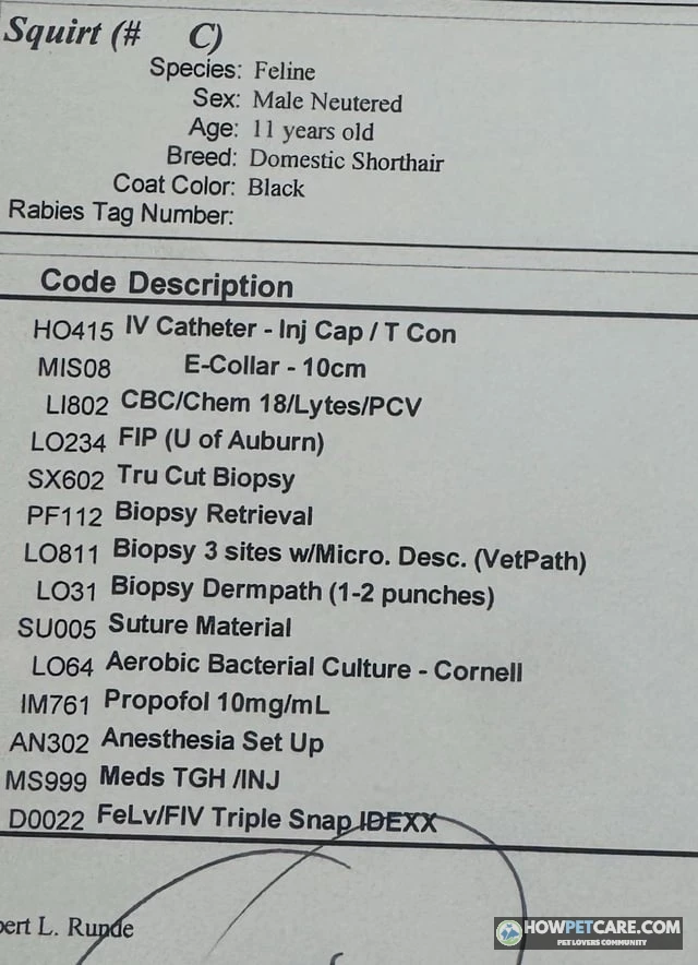
Prices depend on whether you use a general practice, emergency, or specialty clinic; an in-house or reference lab; sedation; the number of nodes sampled; and whether a delayed diagnosis leads to repeat visits.
Which test to do first if money is tight
A reasonable sequence is exam, then FNA with cytology, then FeLV/FIV, then bloodwork, with biopsy and imaging reserved for unclear results or suspected cancer. Skipping the FNA can be a false economy: an unsampled node can lead to repeat visits and empirical antibiotic courses that cost more than the needle sample would have.
Request an itemized estimate and ask which tests would change the treatment plan and which can wait. You can also ask whether the clinic will reuse samples or slides if referral is needed, and check veterinary school clinics, community or low-cost clinics, and payment plans. Compare total costs, not just the visit fee. No insurance or financing product is recommended here; the goal is to know the price before you approve it.
Treatment and Outlook: From Antibiotics to Lymphoma Prognosis
Treating reactive and infectious causes
Reactive nodes usually need nothing more than time and treatment of the trigger: the vaccine reaction or mild cold resolves, and the node follows. Warm compresses aren’t a substitute for diagnosis, and they can make an abscess worse. Infections may need antibiotics, abscess drainage, or dental work. When doxycycline is prescribed, a typical dose is about 5 mg per pound per day, and treatment for tissue infections generally runs a minimum of 10 to 14 days. Your vet determines those numbers for your cat. Never start leftover or online antibiotics on your own.
Lymphoma treatment and survival expectations
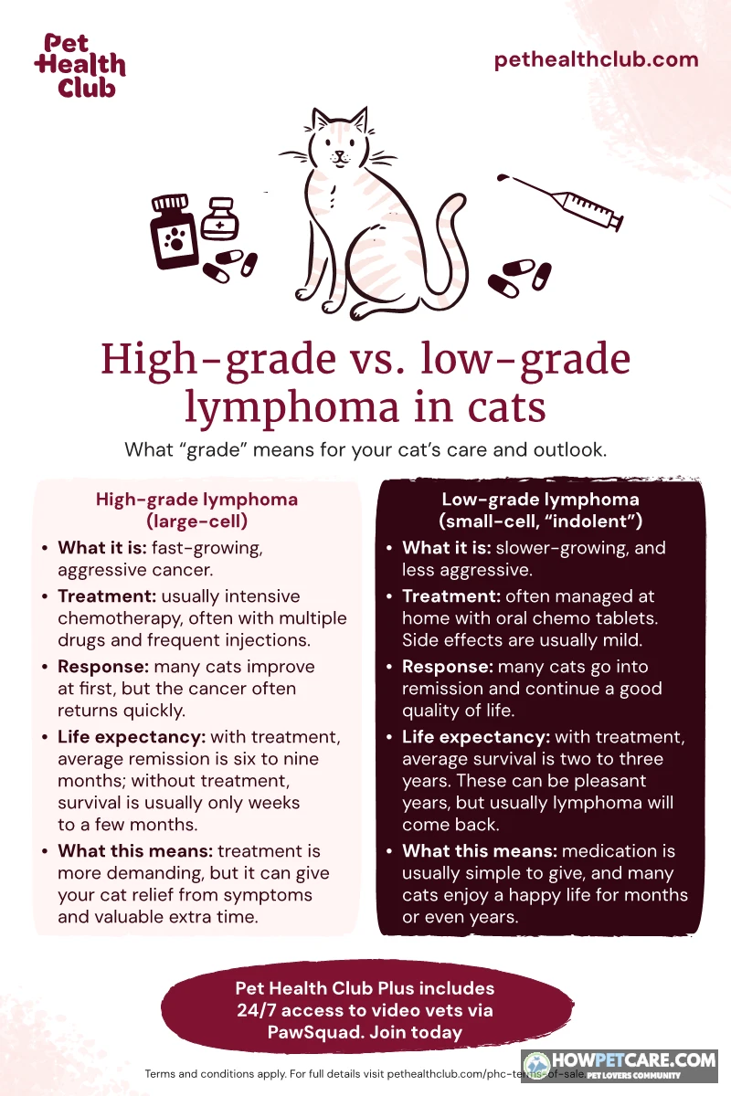
Treatment options include multi-drug chemotherapy protocols, single-agent chemotherapy, and steroids for comfort. Outcomes depend on type, location, grade, and FeLV status.
The figures below are median survival times commonly cited in veterinary oncology literature (published COP/CHOP protocol studies and small-cell intestinal lymphoma cohorts). A median means half of cats live shorter and half live longer — your cat is not a statistic.
| Scenario | Median survival (approximate) |
|---|---|
| Untreated high-grade lymphoma | ~1–2 months |
| Steroids (prednisolone) alone — palliative | ~1–3 months |
| Chemotherapy (CHOP-type protocol), high-grade | ~6–9 months; some cats reach 12+ months |
| Small-cell (low-grade) intestinal lymphoma, chlorambucil plus steroid | Often 2+ years; frequently managed like a chronic disease |
| Kidney or nervous-system lymphoma | Generally more guarded than other forms |
What changes these numbers: FeLV status (FeLV-positive cats with lymphoma historically have shorter remissions and more complications); type and location (intestinal small-cell carries the best outlook); response to the first treatment; and how well the cat feels day to day. Ask your vet or a veterinary oncologist for the range specific to your cat’s diagnosis, cell type, and staging before deciding anything.
FeLV and FIV outlook
FIV-positive cats can live for years with supportive care and monitoring. FeLV prognosis is generally more guarded, and the stage at diagnosis and any co-infections affect the outcome. For both viruses, keep your cat indoors, away from other cats’ infections, and on a consistent preventive care schedule. See the vet sooner rather than later when something changes.
Comfort care and nutrition for serious diagnoses
For a cat with a terminal diagnosis, palliative care focuses on pain control, appetite support, hydration, soft bedding, easy litter access, and reducing stress. A simple quality-of-life check covering eating, drinking, grooming, mobility, interest in interaction, and comfort can help you judge whether your cat is still enjoying life. Track it every few days; hospice and palliative-focused veterinarians can help with the assessment. During treatment, nutrition means a highly palatable, balanced diet and vet-guided appetite stimulation rather than supplements marketed for "immune support." Those supplements lack strong evidence and should be cleared with your vet first.
Acetaminophen and ibuprofen are toxic to cats, and leftover antibiotics can mask the cause and delay diagnosis. Only give what your vet has prescribed for this cat.
1A cat owner notices a single swollen lymph node under their cat's jaw that has been present for three weeks. The cat continues to eat, drink, groom, and play normally, with no fever or signs of distress. How should this scenario be triaged?
Nodes that stay enlarged beyond 7–14 days, even in a cat exhibiting normal behavior, require professional evaluation and sampling to rule out underlying infection or neoplasia.
2An owner notices a lump in their cat's neck and jaw region. Which of the following clinical findings strongly suggest a tooth-root abscess or dental infection rather than a primary swollen lymph node?
Dental abscesses originate at the tooth root fixed within the bone, whereas lymph nodes sit in soft tissue and typically retain some mobility unless fixed by advanced pathology.
3When diagnostic funds are limited, which initial test offers the highest diagnostic yield per dollar for evaluating a persistent, enlarged feline lymph node?
FNA with cytology is low-cost, minimally invasive, usually requires no sedation, and rapidly distinguishes between reactive tissue, active infection, and cellular markers of lymphoma.
4A veterinarian detects an enlarged popliteal lymph node during a physical examination of a cat. Which region of the body should be inspected first for a underlying wound or infection?
The popliteal lymph node is located behind the knee joint and specifically drains the tissue and skin of the lower hind limb.
5Which of the following physical characteristics and clinical patterns are typical of node enlargement caused by feline lymphoma rather than an acute bacterial infection?
Malignant nodes often display firm texture, reduced mobility relative to surrounding tissue, and persistent growth.
6An owner is monitoring a mild, soft lymph node enlargement that appeared after their cat's routine vaccination. If the owner sets a reminder for day 14, which observation requires scheduling a routine veterinary appointment?
Benign reactive swelling should show clear signs of regression by day 14; static size, enlargement, or increased firmness signals the need for diagnostic evaluation.
7What is the primary reason why caretakers are advised to use a humane box trap rather than bare hands when capturing a feral cat with a swollen lymph node?
Direct handling of unsocialized cats poses high human bite/scratch injury risks and potential transmission of infectious agents like Bartonella (cat scratch disease).
8A cat diagnosed with small-cell (low-grade) intestinal lymphoma is managed with chlorambucil and a steroid regimen. Based on published veterinary oncology studies, what is the expected median survival outcome for this specific form of cancer?
Small-cell (low-grade) intestinal lymphoma carries a significantly better prognosis than high-grade forms, with median survival frequently exceeding two years under appropriate medical management.
Swollen Lymph Node in a Stray or Feral Cat When You Can’t Get to a Vet
Observe from a distance and write down whether the cat is eating, moving normally, breathing comfortably, and maintaining body weight, as well as whether the swelling is growing. Take dated photos on a schedule so you can compare them rather than rely on memory. Don’t try to palpate an unsocialized or frightened cat: bites and scratches can cause injury and transmit Bartonella.
For capture, use a humane box trap or a carrier with a towel over it, never bare hands. Community resources can help: try trap-neuter-return groups, humane societies, low-cost and community clinics, and rescue networks. Many test for FeLV/FIV at a modest fee. Signs that justify urgent trapping include labored breathing, a rapidly growing or draining swelling, inability to eat, or severe lethargy.
Don’t give human medications, medicated food, or antibiotics without a diagnosis, and keep the cat separated from your own cats if FeLV is a possibility. If the cat is declining and no care is available, talk to a rescue or a veterinarian about humane options rather than letting the condition go untreated.
Your Next Steps: A Checklist to Triage, Record, and Prepare for the Vet
-
Check for red flags
Use the three-tier list at the top of this guide. The first box you tick sets your deadline.
-
Palpate and record
Check all five reachable node locations, compare left to right, and log location, size versus a familiar object, texture, mobility, and date. Photograph the lump with a coin for scale.
-
Look for lookalikes and triggers
Note anything that fits: bad breath, drooling, a fight wound, a recent vaccine, or a cold that’s going around.
-
Set a date
Choose an emergency clinic today for red flags, a routine visit within a few days for a persistent or growing node, or a phone reminder for day 14 if you’re monitoring a mild case.
Bring the dated photos, your symptom log, vaccine and flea records, and a list of current medications to the appointment. Prepare four questions: which nodes are enlarged, can we do an FNA today, what will each test cost, and which tests can wait?
For red flags, seek same-day emergency care. Call your regular vet within a few days for anything persistent, growing, firm, or paired with other symptoms. Re-check in one to two weeks only if the node is small and soft and your cat is fully normal otherwise.
Most swollen nodes have a treatable cause. An early needle sample is the fastest, cheapest way to find out which kind you’re dealing with.
| Cause Category | Typical Size | Pain on Touch | Warmth | Mobility under Skin | Number Affected | Duration | Accompanying Symptoms | Common Triggers |
|---|---|---|---|---|---|---|---|---|
| Reactive / benign | Pea to small grape; mild enlargement | Usually not painful | Normal temperature | Soft, slides freely | Usually one node near the trigger (e.g., jaw node after dental work) | Days to ~2–4 weeks, then shrinks | Recent vaccine, skin wound, dental disease | Vaccination, mild URI, flea allergy, dental inflammation |
| Infectious | Pea to walnut; can be quite large | Often painful, especially with abscess | Often warm or hot | Movable early; may stick if abscessing | One or a few near the infection site | Persists until infection is treated | Fever, discharge, lethargy, fight wounds, poor appetite | URI, bite abscess, Bartonella (cat scratch), fungal infection, FIV/FeLV-related infection |
| Cancerous (e.g., lymphoma) | Pea to golf ball; keeps growing | Usually not painful — a painless growing node is the concerning pattern | Usually normal temperature | Often firm, may feel fixed or "stuck" | Often several nodes at once, in different body areas | Weeks to months, progressively enlarging | Weight loss, appetite change, possibly vomiting/diarrhea; cat may seem "off" without a clear infection | Feline lymphoma; cancer spread from another site; FeLV raises lymphoma risk |
FAQ
Can swollen lymph nodes in cats go away on their own?
Yes, reactive nodes caused by a vaccine, a minor wound, or a mild infection often shrink within a couple of weeks once the trigger resolves. A node that stays enlarged, keeps growing, or feels firm should be sampled by a vet, because a cause such as lymphoma will not resolve by itself.
Can I give my cat human medication for a swollen lymph node?
No. Common human pain relievers such as acetaminophen and ibuprofen are toxic to cats, and leftover antibiotics can hide the cause and delay diagnosis. Only give medication that your vet has prescribed for your cat.
What other symptoms come with swollen lymph nodes in cats?
Depending on the cause, you may see fever, lethargy, reduced appetite, weight loss, vomiting or diarrhea, nasal or eye discharge, bad breath, or drooling. Several of these together with a swollen node make a vet visit more urgent.
Are swollen lymph nodes in cats contagious?
The swelling itself is not contagious, but some of its causes can spread between cats, including upper respiratory infections and FeLV. Keep a cat with an unexplained swollen node away from other cats until a vet has tested for FeLV and FIV.
Can stress or anxiety cause a cat’s lymph nodes to swell?
Stress alone is not a recognized direct cause of enlarged lymph nodes. Stress can make infections more likely, so a swollen node in a stressed cat still needs to be checked for an underlying cause.