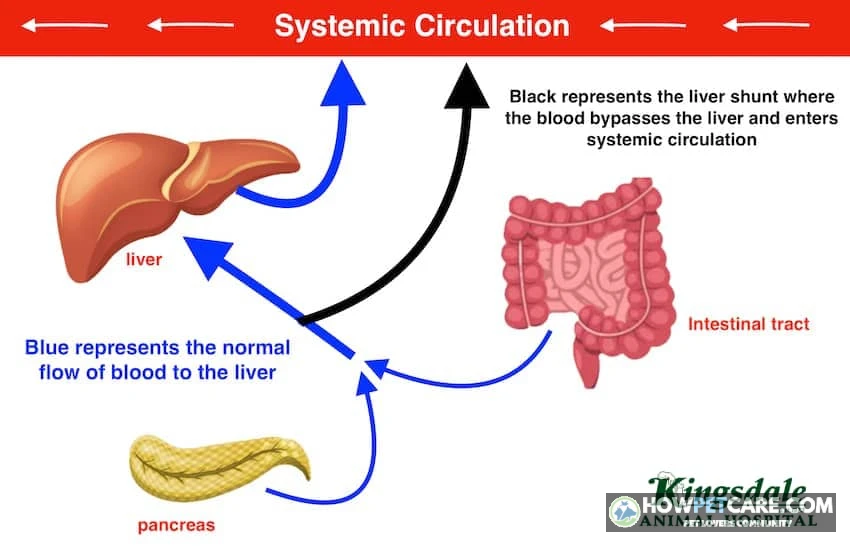

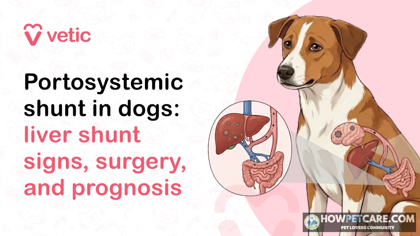
A liver shunt in dogs (portosystemic shunt) is an abnormal blood vessel that diverts blood around the liver instead of through it. Unfiltered toxins such as ammonia reach the brain, causing confusion, seizures, poor growth, and urinary problems. Diagnosis requires veterinary testing; treatment may involve diet, medication, or surgery.
Liver Shunt in Dogs: What It Is and the Red Flags That Need a Vet Now
A liver shunt in dogs is an abnormal blood vessel that allows blood to bypass the liver instead of passing through it for processing. Waste products, including ammonia, can then reach the brain and cause confusion, abnormal behavior, or seizures. A dog with a suspected liver shunt needs veterinary testing. Treatment may involve medication, a carefully chosen diet, or a procedure to reduce blood flow through the vessel.
Many dogs can be helped. The right treatment and outlook depend on the type of shunt, the dog’s condition, and whether the vessel can be closed safely.
The 30-second explanation
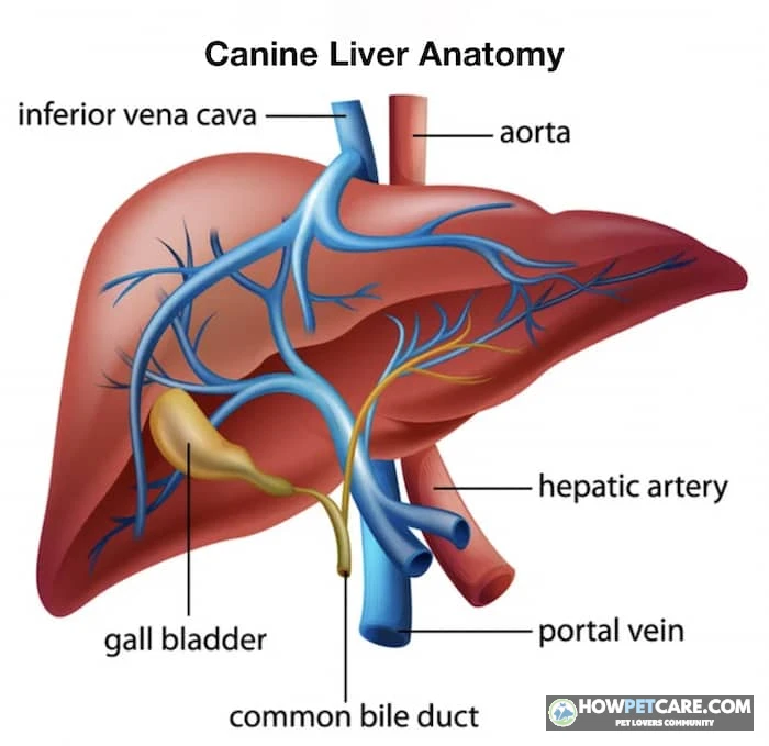
Normally, blood carrying nutrients and other substances from the digestive tract passes through the liver before circulating around the body. A portosystemic shunt creates a detour around the liver. The resulting buildup of substances that affect the brain is called hepatic encephalopathy. Dogs may also have digestive or urinary problems and, particularly as puppies, grow poorly.
Red-flag checklist: when to go to the vet or ER immediately
Go to an emergency veterinarian now if your dog has:
- A seizure or convulsions, even if the episode appears to have stopped.
- Collapse, unresponsiveness, or an inability to stand.
- Sudden blindness, head pressing, severe disorientation, persistent circling, or rapidly worsening behavior after a meal.
- Repeated or prolonged vomiting or diarrhea, especially with weakness, lethargy, or other neurological signs.
- Straining to urinate with little or no urine produced. A urinary blockage is an emergency, whether or not a shunt is involved.
Call the clinic while you’re on the way, if possible. During a seizure, keep your dog away from stairs and hard objects; don’t put your hands in the dog’s mouth.
Call your regular veterinarian promptly about milder signs, such as brief episodes of staring after meals, poor puppy growth, increased thirst or urination, blood in the urine, or intermittent digestive upset. Ask how soon your dog should be examined. If neurological signs recur, persist, or worsen, seek urgent care rather than waiting for a routine appointment.
A symptom checklist can’t diagnose or rule out a liver shunt. Several other conditions can cause similar signs.
Dog Liver Shunt Symptoms: What Owners Notice in Puppies and Adult Dogs
Neurological signs
A dog with hepatic encephalopathy may seem unusually quiet or confused, stare into space, pace, circle, press its head against objects, stumble, or have seizures. Signs can come and go, so a short video may help the veterinary team.
Tell your vet if your dog has had an unusual or prolonged response to a sedative or anesthetic. It’s a clue worth discussing, not proof of a shunt.
Growth, digestive, and urinary signs
Puppies with a liver shunt may be smaller than their littermates or fail to gain weight as expected. Vomiting, diarrhea, poor appetite, and lethargy can also occur. Congenital shunts are present at birth, though signs may not be obvious right away; some dogs aren’t diagnosed until adulthood.
Urinary signs can include increased drinking or urination, blood in the urine, and straining. Some affected dogs develop ammonium biurate bladder stones, which can cause serious urinary problems. If your dog is straining, contact a vet promptly. If little or no urine is coming out, go to an ER.
Why symptoms often worsen after eating
Substances produced during digestion, including ammonia, normally reach the liver for processing. When blood bypasses the liver, neurological signs may become more noticeable after a meal. Not every dog follows this pattern, so normal behavior after eating doesn’t rule out a shunt.
Record what happened, when your dog last ate, how long the episode lasted, and whether your dog recovered fully. Don’t delay an urgent visit to collect a longer log.
Types and Causes: Congenital vs Acquired, Intrahepatic vs Extrahepatic Shunts
Congenital vs acquired
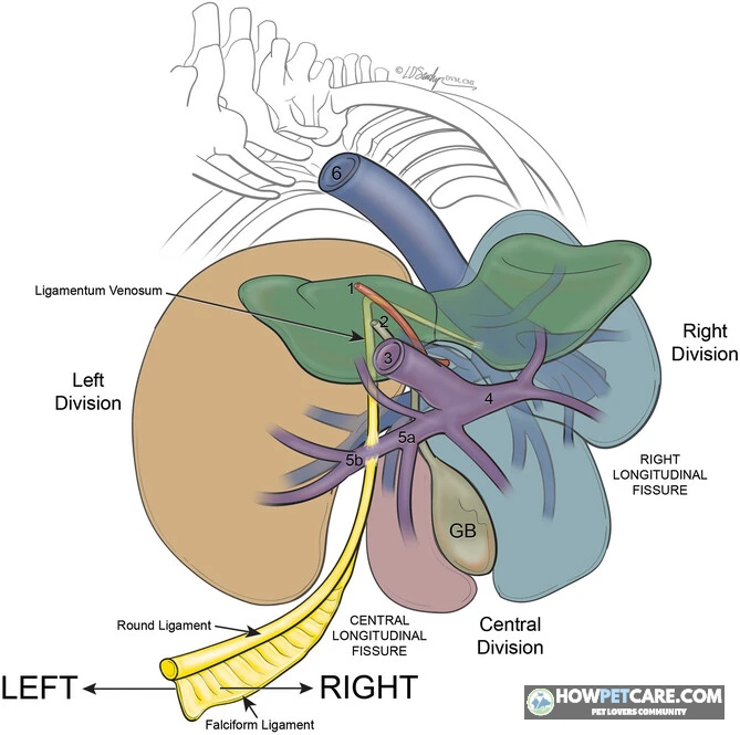
A congenital liver shunt is present at birth. Breed predispositions suggest a hereditary component in some dogs. Many congenital shunts involve a single abnormal vessel that a specialist can assess for closure.
Acquired shunts develop later, usually when chronic liver disease causes high pressure in the portal circulation, known as portal hypertension. These shunts often consist of multiple vessels that form alternative routes for blood flow. Closing them doesn’t correct the underlying high pressure, so treatment generally focuses on the liver disease and its consequences rather than closing the vessels.
Extrahepatic vs intrahepatic
These terms describe the vessel’s location:
- Extrahepatic shunt: The abnormal vessel runs outside the liver. These shunts are more typical of small and toy breeds and are often more accessible for surgical treatment.
- Intrahepatic shunt: The vessel runs within the liver. These shunts occur more often in larger breeds and can be more technically challenging to treat.
Location is only part of the decision. A specialist must also assess the number and connections of vessels, the liver’s blood supply, and whether the dog can tolerate a procedure.
Liver shunt in small-breed dogs and at-risk breeds
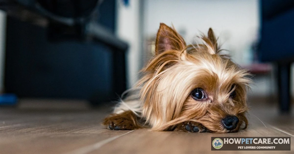
Yorkshire Terriers and Maltese are among the small breeds associated with congenital shunts. Miniature Schnauzers are also among the predisposed breeds, while Irish Wolfhounds are among the larger breeds affected. Mixed-breed dogs can have shunts too; breed alone can’t confirm or exclude one.
How a Canine Portosystemic Shunt Is Diagnosed: Tests, What They Show, and Typical Costs
Step-by-step diagnostic flow
Your veterinarian will start with your dog’s history and an examination, then select tests to look for evidence of abnormal liver function and identify the vessel. Not every dog needs every test, and the order may change if your dog is unstable. A veterinary internal medicine specialist or surgeon may coordinate advanced imaging and treatment planning.
Bile acid and ammonia tests
A bile acid test often compares samples taken before and after a meal. High bile acids can indicate that blood is bypassing the liver or that the liver isn’t functioning normally; the result alone doesn’t identify the cause. Ammonia levels can provide useful evidence, but the sample needs careful handling. The result must be interpreted alongside the examination and other tests.
Ultrasound and CT angiography
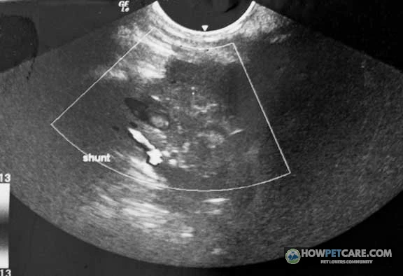
An experienced clinician may use ultrasound to look for an abnormal vessel and assess the liver and urinary tract. CT angiography provides a detailed map of blood vessels and is particularly useful for planning treatment. Your specialist can explain which imaging study is appropriate for your dog.
| Test | What it shows | Limitations | Typical cost range (estimate) |
|---|---|---|---|
| Examination and history | Growth, neurological signs, hydration, and symptom pattern | Cannot confirm a shunt | Ask the clinic for its exam fee |
| Blood count, chemistry, and urinalysis | Clues such as low blood urea nitrogen, small red blood cells, or urinary crystals | Findings are not specific to shunts and may not all be present | Request an itemized laboratory estimate |
| Bile acid test | Whether bile acids rise abnormally, suggesting impaired liver processing or bypass | Other liver problems can also cause high results | Ask whether both samples and lab fees are included |
| Ammonia test | Whether ammonia is elevated | Sample handling matters; results do not map the vessel | Ask about collection, processing, and laboratory fees |
| Abdominal ultrasound | May show a shunt and other abdominal abnormalities | Results depend on the dog and the operator; a vessel may be difficult to visualize | Request the imaging and specialist-reading fees |
| CT angiography | Maps vascular anatomy for diagnosis and procedure planning | Requires specialist equipment and usually anesthesia or sedation | Ask for the scan, contrast, anesthesia, and interpretation costs |
The supplied evidence doesn’t establish reliable national prices for these individual tests. Fees vary substantially, so a written local estimate is more useful than treating an unsourced figure as a standard charge.
Medical Management vs Surgery: How to Choose for Your Dog
When medical management makes sense
Medical care is often used to stabilize a dog before a procedure. It can also be the longer-term plan when surgery isn’t feasible, when the shunt is acquired, or when finances, specialist access, or other health problems limit treatment choices.
A veterinarian may recommend a controlled-protein diet, lactulose to help reduce absorption of ammonia-producing substances, and antibiotics in selected cases. Seizure treatment may also be needed. These measures aim to control signs; they don’t close the abnormal vessel. Some dogs maintain a good quality of life with medical care, while others continue to have episodes or deteriorate.
When surgery is usually recommended
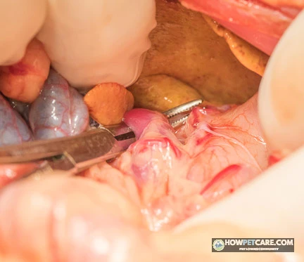
For a suitable dog with a single congenital shunt, a specialist may recommend a procedure to reduce or close the abnormal blood flow. The goal is to direct more blood through the liver without raising pressure in the portal circulation to a dangerous level.
The choice isn’t simply surgery versus doing nothing. Ask the team to weigh the likely benefit of intervention against your dog’s current stability, shunt anatomy, other illnesses, access to a qualified center, and your ability to manage follow-up care. Age alone doesn’t make the decision.
Questions to ask your surgeon before deciding
Ask which vessel has been identified, whether it appears congenital or acquired, and whether the team expects it can be safely narrowed or closed. Discuss the proposed technique, the surgeon’s experience, possible complications, the hospital stay, and the backup plan if complete closure isn’t possible.
Liver Shunt Surgery Options Compared: Ameroid Constrictor, Cellophane Band, Ligation, and Coil Embolization
Extrahepatic shunt surgery
For a suitable vessel outside the liver, a surgeon may choose a method intended to narrow blood flow gradually:
- Ameroid constrictor: A device placed around the vessel and designed to narrow its opening over time.
- Cellophane band: A band placed around the vessel to encourage gradual narrowing as tissue responds.
- Suture ligation: A surgical tie used to narrow the vessel. The surgeon determines how much closure the dog can tolerate; complete closure isn’t always safe at the first procedure.
Gradual narrowing gives the liver and portal circulation time to adapt, but neither a device nor a tie guarantees complete closure. The best choice depends on the anatomy and the surgical team’s assessment.
Intrahepatic shunt treatment
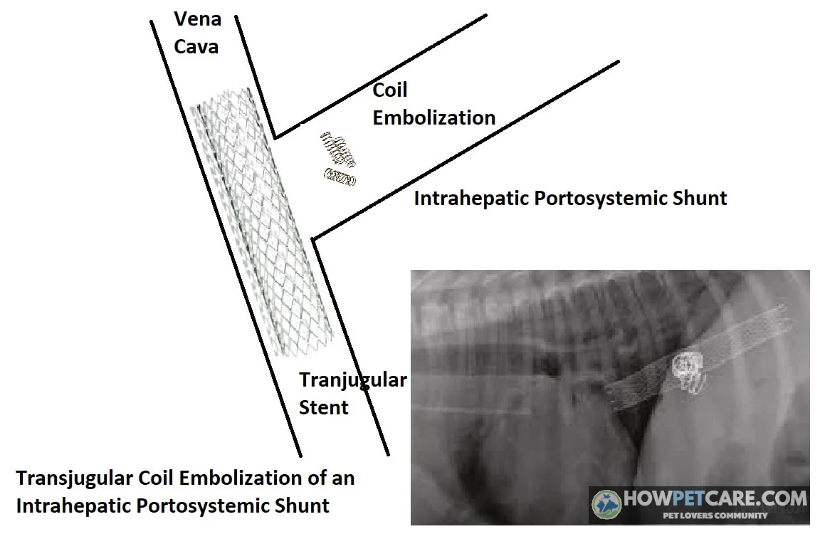
For some shunts within the liver, a specialist may offer coil embolization through interventional radiology. The team accesses the circulation with a catheter and places devices to reduce flow through the shunt, often using a stent as part of the procedure. This approach avoids an open abdominal operation, but it requires specialized equipment and expertise. Other intrahepatic shunts may be managed surgically or medically.
Risks and complications
Possible complications include dangerously increased portal pressure, seizures after a procedure, persistent shunting, and the development of additional abnormal vessels. A dog may need continued medication, repeat imaging, or further treatment. Ask what signs the hospital monitors for and what its plan is if a complication occurs.
The evidence supplied for this article doesn’t establish comparable success or complication percentages for each technique. Ask the treating center about outcomes for dogs with anatomy similar to your dog’s and how it defines a successful result.
Liver Shunt Surgery Cost for Dogs: Full Budget From Testing to Aftercare and Insurance
Itemized cost breakdown
A procedure quote is only part of the budget. Ask each provider for a written low-to-high estimate and confirm what’s included before adding figures together. The table is a worksheet, not a national fee schedule. The supplied evidence doesn’t verify current prices for these services, so presenting precise ranges as established U.S. rates would be misleading.
| Stage | What is included | Typical cost range (estimate) | Notes and variables |
|---|---|---|---|
| Initial workup | Exam, bloodwork, urinalysis, bile acids, and sometimes ammonia | Request a written range | Tests may be staged rather than performed together |
| Advanced imaging | Ultrasound, CT angiography, specialist interpretation, and possible anesthesia | Request separate ultrasound and CT ranges | Some dogs need both; ask about contrast and anesthesia |
| Emergency stabilization | ER examination, monitoring, fluids, and seizure or encephalopathy treatment when needed | Request a range for the expected stay | An emergency or extended hospitalization can substantially increase the bill |
| Surgery or catheter procedure | Specialist fees, operating facilities, devices, and anesthesia | Request a procedure-specific low and high estimate | Location, technique, hospital, and complications affect cost |
| Hospitalization | Nursing, monitoring, medications, and inpatient care | Ask whether included in the procedure quote | Extra nights or intensive care may be billed separately |
| Follow-up | Recheck visits, bloodwork, bile acids, and imaging if indicated | Request a follow-up plan and estimate | Tests depend on recovery and the specialist’s assessment |
| Medications | Lactulose, antibiotics, or seizure medication when prescribed | Ask for a monthly estimate | Cost changes with the dog’s size, drugs, and treatment duration |
| Diet | Veterinary-recommended hepatic or other suitable food | Ask for a monthly estimate | Food size, formula, and daily intake matter |
To compare your options, add the initial workup, imaging, procedure, any separately billed hospitalization, and planned follow-up. Then add a contingency for complications. For medical management, add the initial workup to the recurring food, medication, and monitoring estimates; ask how often rechecks are expected. Avoid counting an imaging study or hospital stay twice if it’s already included in the specialist’s package.
Will pet insurance cover a liver shunt?
Coverage depends on the policy and your dog’s history. Some plans cover eligible congenital or hereditary conditions, while others exclude them or impose conditions. Signs, testing, or a diagnosis documented before coverage or during a waiting period may be treated as pre-existing, even if the shunt was confirmed later.
Ask the insurer for a written coverage determination where available. Check the waiting period, hereditary and congenital-condition language, annual limit, deductible, reimbursement rate, and whether examinations, imaging, prescription food, and complications are covered.
Ways to manage the cost
Ask the hospital whether it offers staged estimates, payment arrangements, or veterinary financing. A university hospital, local nonprofit, or charitable fund may be able to suggest assistance, though availability and eligibility vary. If the full plan is unaffordable, tell the veterinarian early. They can discuss which tests are most useful now and what a medically managed plan would involve.
Life Expectancy and Prognosis: Surgical vs Medical Management by Shunt Type
Side-by-side outcomes
There is no single life expectancy for a dog with a liver shunt. Outcomes differ with anatomy, underlying liver health, complications, and how completely blood flow through the shunt can be reduced.
| Shunt type / approach | Common treatment | Main benefits | Main risks | Reported outcomes (with source) |
|---|---|---|---|---|
| Single congenital extrahepatic shunt | Surgical narrowing or closure when feasible | Can redirect blood toward the liver and substantially improve signs | Portal hypertension, seizures, or persistent shunting | Small-breed extrahepatic shunts are generally more accessible to correction than intrahepatic shunts; the supplied evidence does not establish a survival percentage |
| Single congenital intrahepatic shunt | Catheter-based embolization or surgery at a specialist center; medical care when intervention is unsuitable | Can reduce abnormal flow; catheter treatment may avoid open abdominal surgery | Technical difficulty, complications, or residual shunting | Intrahepatic shunts are generally more challenging to correct; a comparable survival percentage is not established in the supplied evidence |
| Congenital shunt managed medically | Diet and prescribed medication, with monitoring | Can control signs and support quality of life without a procedure | The vessel remains open; episodes may recur or progress | Individual responses vary; the supplied evidence does not support a numerical survival comparison |
| Acquired multiple shunts | Management of underlying liver disease and complications | Addresses the disease driving abnormal blood flow | Prognosis depends on the underlying disease and portal hypertension | Acquired shunts are associated with chronic liver disease and portal hypertension; congenital-shunt surgical statistics should not be applied to them |
What affects an individual dog’s outlook
Can a liver shunt in dogs be cured? Some congenital shunts can be closed successfully, with major improvement or resolution of signs. Others can’t be fully closed safely, and some dogs need ongoing treatment or further assessment. A successful operation doesn’t eliminate the need for recommended rechecks.
If your dog is managed medically or recovering from a procedure, track appetite, weight, muscle condition, energy, sleep, urination, and the frequency and severity of neurological episodes. Share any changes with the veterinary team. Ask what improvement is realistic for your dog and what changes would prompt a different treatment plan.
Diet for Dogs With a Liver Shunt: Protein Targets, Sources, and Prescription Options
Protein targets and why restriction needs supervision
Diet can help reduce substances that contribute to hepatic encephalopathy, but too little protein can cause muscle loss and is especially concerning in a growing puppy. A low-protein diet isn’t automatically right for every dog with a suspected shunt.
Ask your veterinarian for an individualized protein target based on your dog’s age, growth, body and muscle condition, neurological signs, and overall nutritional needs. No single protein percentage is appropriate for every patient. A veterinary nutrition specialist can help when a puppy needs a growth-appropriate plan or a dog won’t eat a suitable commercial food.
Best protein sources and prescription diet examples
Some dogs tolerate diets using dairy or plant-based proteins, such as soy, better than diets relying heavily on certain meat proteins. Tolerance varies, and the complete diet matters more than a single ingredient. Ask your vet whether a veterinary hepatic-support formula, such as Hill’s Prescription Diet l/d or Royal Canin Hepatic, is appropriate and available for your dog’s life stage.
Don’t switch a puppy to an adult prescription food or prepare a restrictive homemade diet without veterinary guidance. Treats, chews, table scraps, and supplements count as part of the diet too; bring their labels to the appointment.
Daily feeding routine
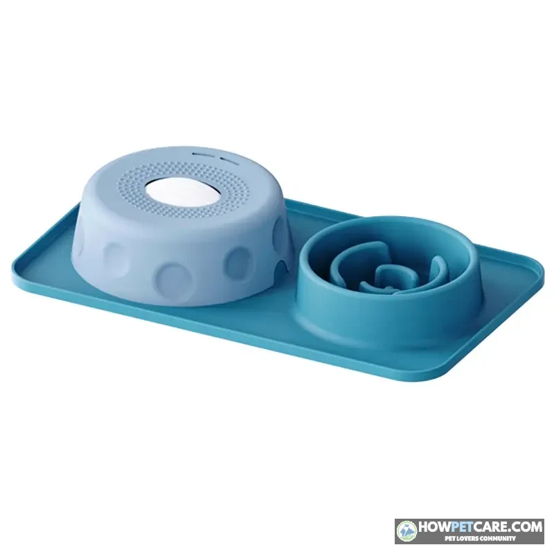
Your vet may suggest smaller, more frequent meals and a consistent feeding routine. Keep fresh water available, measure the prescribed food, and monitor weight and muscle condition. Diet works alongside—not in place of—prescribed lactulose or other medication. Ask when to call if your dog refuses food, vomits, develops diarrhea, or shows signs after eating.
Do not sharply reduce protein, stop prescribed medication, or give supplements marketed for liver detoxification without checking with your veterinarian. A growing puppy needs a nutritionally appropriate plan.
Recovery, Follow-Up Testing, and Breeding: What Happens After Diagnosis
Week-by-week recovery timeline
Recovery varies by procedure and dog. Follow the hospital’s discharge instructions rather than a generic calendar.
- Hospital stay: The team monitors neurological status, pain, appetite, and signs of increased portal pressure. The length of stay depends on the procedure and recovery.
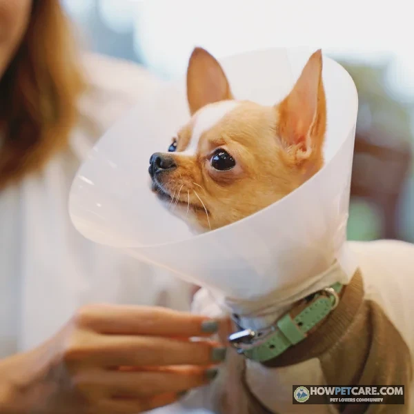
- First days at home: Give medications exactly as directed. Check any incision as instructed, prevent licking, and follow the activity and feeding plan. Don’t discontinue lactulose or change the diet just because your dog seems brighter.
- Following weeks: Return for the scheduled examination and any incision check. Gradually resume activity only when the surgeon says it’s safe. Record appetite, bowel movements, energy, and any unusual behavior.
- Later recovery: The specialist assesses whether signs and test results have improved and whether medication or food can be adjusted. Some dogs need longer-term treatment.
Contact the surgical team promptly for worsening pain, abdominal swelling, repeated vomiting, marked lethargy, or incision discharge. Seizures, collapse, severe disorientation, or inability to stand warrant emergency care.
Follow-up bloodwork and imaging
Your surgeon may recommend repeat bloodwork and bile acids after recovery, with ultrasound or other imaging if results or signs raise concern about persistent shunting. Timing depends on the closure method and your dog’s progress. Ask which test will help assess the result, when it should be done, and whether it’s included in the estimate.
Screening and breeding considerations
Because some congenital shunts have a hereditary component, affected dogs should not be bred. If you’re choosing a puppy in a predisposed breed, ask the breeder about shunts in the parents, littermates, and close relatives, and ask a veterinarian what screening is appropriate. A breeder may discuss bile acid testing, but a normal test in a parent doesn’t guarantee that a puppy will be unaffected.
Your Next Steps: Pre-Appointment Checklist and Questions to Ask Your Vet
Pre-appointment checklist
Bring a short record of what you’ve seen. You can use this format:
Date and time | Last meal | Signs and duration | Recovery | Video available?
Also bring your dog’s food and treat information, medications and supplements, prior laboratory results, and any known family history. If your dog is currently severely disoriented, seizing, collapsed, or unable to urinate, go to an emergency clinic instead of waiting to complete the record.
If your dog is stable but symptomatic, call your veterinarian for an appointment and ask whether any signs change the urgency. If a shunt has already been diagnosed, request the imaging report and discuss referral. If a procedure is planned, confirm the stabilization plan, written estimate, and emergency contact before going home.
Questions to ask your veterinarian or surgeon
- How confident are you that a shunt is present, and what else could explain these signs?
- Is the suspected shunt congenital or acquired, and is it inside or outside the liver?
- Which tests are most useful next, and do we need ultrasound, CT angiography, or both?
- Should my dog see a veterinary internal medicine specialist or surgeon?
- Is my dog a candidate for a procedure, and which technique do you recommend for this anatomy?
- What complications have you seen with this approach, and what happens if the shunt cannot be fully closed?
- If we manage medically, what signs would show that the plan is or is not working?
- What food and protein target are appropriate for my dog’s age, growth, and muscle condition?
- What does the written estimate include for imaging, anesthesia, hospitalization, medications, and follow-up?
- Which recheck tests will you recommend, and when might medication or diet be adjusted?
- Which changes mean I should call you, and which mean I should go straight to an ER?
- Are payment arrangements or assistance programs available, and what documentation should I send my insurer?
Before leaving, make sure you know whom to call after hours and what to do if your dog has another episode.
1What primary mechanism causes neurological signs (hepatic encephalopathy) in a dog with a liver shunt?
When blood skips liver processing, intestinal toxins accumulate in the systemic circulation and alter brain function.
2Why do neurological symptoms in dogs with a liver shunt often become more noticeable shortly after eating?
Post-meal digestion increases the production of nitrogenous waste in the intestines, which enters blood that bypasses liver detoxifying pathways.
3How do extrahepatic shunts differ from intrahepatic shunts in terms of location and breed tendencies?
Vessel location relative to liver tissue correlates with body size, with small/toy breeds favoring outside vessels and large breeds favoring internal ones.
4Which statement accurately contrasts congenital liver shunts with acquired liver shunts?
Congenital shunts are developmental anomalies of single vessels, while acquired shunts form multiple collateral vessels to relieve high portal blood pressure.
5What is a primary limitation of relying solely on a blood bile acid test when evaluating a dog for a liver shunt?
Bile acid testing measures function/bypass capability; advanced imaging is required to map blood vessel anatomy.
6Why is CT angiography particularly valuable in the diagnostic workup of a dog suspected of having a portosystemic shunt?
CT angiography accurately highlights vessel structure, location, and connections, guiding surgical planning.
7What is the primary objective of medical management (such as lactulose and controlled-protein diets) in dogs with liver shunts?
Medical therapies lower circulating ammonia and symptom frequency, but they do not alter the physical vascular detour.
8Why do surgeons often prefer devices like ameroid constrictors or cellophane bands over immediate complete suture ligation for extrahepatic shunts?
Abruptly closing a shunt can cause life-threatening portal hypertension; gradual occlusion gives the microvasculature time to handle increased flow.
9What is a key feature of catheter-based coil embolization when treating intrahepatic liver shunts?
Coil embolization accesses the vascular system via intravascular catheters to occlude internal shunt vessels minimally invasively.
10Why is unmonitored severe protein restriction dangerous when managing a dog with a suspected or confirmed liver shunt?
While protein yields ammonia, essential amino acids are vital for body mass maintenance and juvenile development.
| Test | What It Shows | Limitations | Typical Cost Range (Estimate) |
|---|---|---|---|
| CT angiography | Maps vascular anatomy for diagnosis and procedure planning | Requires specialist equipment and usually anesthesia or sedation | Ask for the scan, contrast, anesthesia, and interpretation costs |
| Abdominal ultrasound | May show a shunt and other abdominal abnormalities | Results depend on the dog and the operator; a vessel may be difficult to visualize | Request the imaging and specialist-reading fees |
| Bile acid test | Whether bile acids rise abnormally, suggesting impaired liver processing or bypass | Other liver problems can also cause high results | Ask whether both samples and lab fees are included |
| Ammonia test | Whether ammonia is elevated | Sample handling matters; results do not map the vessel | Ask about collection, processing, and laboratory fees |
| Blood count, chemistry, and urinalysis | Clues such as low blood urea nitrogen, small red blood cells, or urinary crystals | Findings are not specific to shunts and may not all be present | Request an itemized laboratory estimate |
| Examination and history | Growth, neurological signs, hydration, and symptom pattern | Cannot confirm a shunt | Ask the clinic for its exam fee |
FAQ
What causes liver shunts in dogs and is it genetic?
Most liver shunts in dogs are congenital, meaning the dog is born with an abnormal blood vessel, and certain breeds have a hereditary predisposition. Acquired shunts develop later from chronic liver disease and high pressure in the portal vein.
Which dog breeds are most prone to liver shunts?
Small breeds such as Yorkshire Terriers, Maltese, Havanese, and Shih Tzus are prone to extrahepatic shunts, while large breeds such as Irish Wolfhounds, Labrador Retrievers, and Golden Retrievers more often have intrahepatic shunts. Any breed or mixed-breed dog can be affected.
Can a dog with a liver shunt be treated without surgery?
Yes, medical management with a controlled-protein diet, lactulose, and antibiotics can control symptoms in some dogs. It does not fix the shunt, so many dogs do better long term with surgery when they are good candidates.
Can a liver shunt in dogs be cured?
Surgery can close or greatly reduce a congenital shunt, and many dogs improve substantially or return to normal. Results depend on shunt type and complications, and some dogs still need follow-up care or medication.
At what age do liver shunt symptoms start?
Most dogs with congenital shunts show signs in the first year of life, often as stunted growth or odd behavior after eating. Dogs with milder shunts may not show clear signs until they are adults.