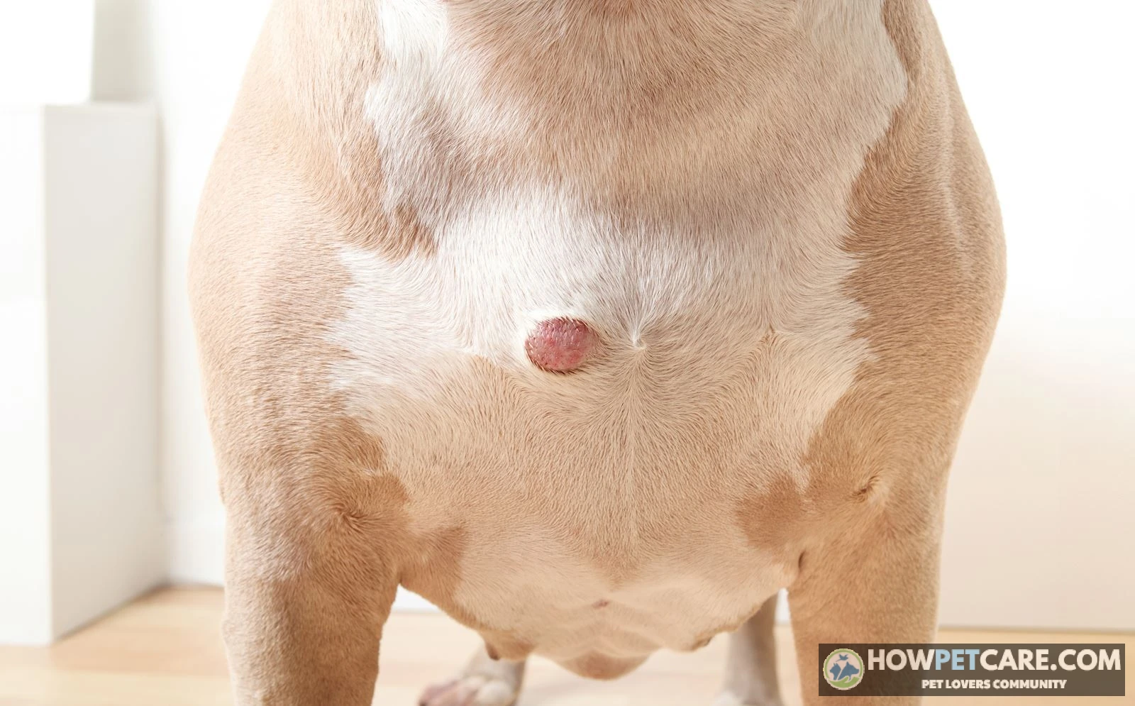

A dog histiocytoma is a benign, rapid-growing skin tumor of Langerhans-cell origin, common in dogs under three years old. Often called a "button tumor," it appears as a raised, hairless red bump that usually plateaus within two weeks and spontaneously regresses without surgery over 8 to 12 weeks via normal immune response.
A canine histiocytoma is a benign cutaneous tumor that almost always resolves on its own. Most young dogs develop a small, red, hair‑less nodule that may ulcerate or bleed, but urgent surgery is rarely needed. The first step is to confirm the diagnosis with a fine‑needle aspiration (FNA) and then follow a structured watch‑and‑wait plan that includes proper wound care and regular veterinary checks. Below you’ll learn how to recognize this “button tumor,” differentiate it from dangerous mimics, understand the natural regression timeline, manage ulceration at home, and budget for every possible treatment option.
What is a canine histiocytoma and why does it appear so quickly?
A histiocytoma is a benign cutaneous tumor of Langerhans‑cell origin that typically shows up as an erythematous, button‑like nodule on the skin. It is the most common skin tumor in dogs under three years of age and often appears within 1–2 weeks after a minor skin irritation or trauma. The rapid onset results from an abrupt proliferation of Langerhans cells in the dermis, part of the immune system that can respond swiftly to local inflammation.

Typical age and breed predispositions
- Age: Most cases occur in dogs younger than 3 years.
- Breeds with higher incidence: Boxer, Great Dane, Dachshund, and Bulldog variants. Some reports also note Shar Pei for the rarer cutaneous Langerhans Cell Histiocytosis, which can behave more aggressively.
Why the tumor surfaces suddenly
The exact trigger remains unclear, but minor scratches, insect bites, or allergic skin reactions can stimulate Langerhans cells to multiply excessively. Because the immune system can expand these cells quickly, the lump becomes visible in just a few days. Growth typically plateaus after 1–2 weeks, helping distinguish it from many malignant skin cancers that keep expanding.
How veterinarians differentiate a benign histiocytoma from mast cell tumors, melanomas, or histiocytic sarcomas
Visual inspection alone cannot rule out a malignant mast cell tumor (MCT) or melanoma. Veterinarians rely on fine‑needle aspiration (FNA) cytology as the first diagnostic step. The aspirated cells are examined under a microscope:
- Histiocytoma cells are large, pale‑staining with characteristic “coffee‑bean” nuclei.
- Mast cells contain dense, purple‑staining granules that appear metachromatic on Wright‑Giemsa stain.
- Melanoma cells are spindle‑shaped, may contain pigment granules, and show prominent nucleoli.
If the cytology is inconclusive, a punch or excisional biopsy provides a larger tissue sample for definitive histopathology.
Never rely on appearance alone; an FNA rules out mast cell disease, which can be life‑threatening.
Clinical Triage Matrix: Histiocytoma vs. High‑Risk Mimics
| Diagnostic Vector | Cutaneous Histiocytoma (Benign) | Mast Cell Tumor (MCT) | Amelanotic Melanoma |
|---|---|---|---|
| Typical Age & Signalment | Dogs < 3 yr; sudden onset | Any age, median 8–9 yr; common in Boxers, Pugs, Labs | Usually older dogs (9–11 yr) |
| Physical Feel & Appearance | Raised, firm, hairless, dome‑/"button" shape; bright pink/red edge | Variable: soft‑squishy to firm, may be pigmented or ulcerated | Flesh‑toned to pale pink, firm, often fast‑growing |
| Growth Dynamics | Erupts 7–14 days, plateaus < 2 cm, then regresses | Continuous growth, may wax/wanes with degranulation | Steady progressive growth, no plateau |
| Darier’s Sign | Negative (no acute wheal on pinching) | Positive (pinch → swelling, redness) | Negative |
| FNA Cytology Hallmarks | Large pale Langerhans cells, coffee‑bean nuclei | Round cells with dense metachromatic granules | Spindle/epithelioid cells, pigment granules, prominent nucleoli |
| Primary Danger | Secondary bacterial infection, misdiagnosis | Systemic anaphylactoid reactions, local invasion | High metastatic potential |
Step‑by‑step FNA procedure
- Restrain the dog and clip a small area of hair over the nodule.
- Disinfect the skin with 70 % isopropyl alcohol.
- Insert a 22‑gauge needle attached to a 1 ml syringe; apply gentle negative pressure.
- Aspirate a few drops of cellular material and expel onto a glass slide.
- Air‑dry the slide, stain (e.g., Diff‑Quik), and hand to the clinic’s pathologist.

Key cytology findings
- Histiocytoma: Large cells, pale cytoplasm, indented nuclei.
- Mast cell tumor: Small to medium cells, abundant granules that stain metachromatically.
- Melanoma: Dark pigment granules, spindle morphology.

When a biopsy is recommended
- Inconclusive or equivocal FNA results.
- Rapid enlargement after the 4‑week plateau.
- Persistent ulceration with foul odor.
- Owner desire for definitive diagnosis before deciding on surgery.
Regression timeline: week‑by‑week stages and at‑home wound care for ulcerated or bleeding tumors
The natural course of a histiocytoma spans 8–12 weeks in most dogs. Understanding each stage helps you recognize normal healing versus a complication that needs veterinary attention.
4‑Stage Spontaneous Regression Roadmap
- Stage 1 – Eruption (Weeks 1–2)
- Visual: Rapidly growing, smooth, hairless, cherry‑pink button (0.5–2 cm).
- Biology: Langerhans cells proliferate after local irritation.
- Care: Schedule FNA; avoid picking or applying any salve.
- Stage 2 – Plateau & Desquamation (Weeks 3–5)
- Visual: Growth stops; surface becomes dull red, may develop a thin central crust.
- Biology: Cytotoxic T‑cells recognize tumor antigens, halting proliferation.
- Care: Measure diameter weekly; keep crust intact.
- Stage 3 – Ulceration & Crusting (Weeks 6–8)
- Visual: Center ulcerates, oozes clear fluid, forms a scab; lesion may appear worse before it gets better, sometimes resembling acute hot spots on dogs due to intense focal inflammation.
- Biology: Immune‑mediated cytolysis destroys tumor cells, causing controlled tissue breakdown.
- Care: Clean twice daily (see cleaning protocol below) and fit an e‑collar.
- Stage 4 – Involution (Weeks 9–12)
- Visual: Nodule flattens, scab dries and falls off, leaving a smooth, lightly pigmented patch that eventually re‑epithelializes.
- Biology: Macrophages clear debris; epidermal cells migrate to restore skin integrity.
- Care: Discontinue routine washes; monitor for hair regrowth.

Weekly visual checklist
| Week | Size (mm) | Surface | Color | Crust/Ulcer? | Red‑flag signs |
|---|---|---|---|---|---|
| 1‑2 | 5‑20 | Smooth, hairless | Bright pink/red | None | Sudden increase > 1 cm |
| 3‑4 | Stable | Dull red, slight scaling | Pink‑purple | Thin dry crust possible | Rapid growth resumes |
| 5‑7 | Stable or slight shrink | Ulcerating, serous fluid | Darker red | Moist crust, possible hemorrhage | Thick purulent discharge, foul odor |
| 8‑12 | Decreasing | Flat, scar‑like | Light brown/gray | Scab falls off | Persistent pain, swelling beyond margins |
Cleaning agents: what to use and what to avoid
- Safe: Sterile 0.9 % saline solution or 0.05 % diluted chlorhexidine diacetate (sky‑blue tint). Apply with a sterile gauze pad; do not scrub.
- Avoid: Hydrogen peroxide (damages healthy tissue), rubbing alcohol (painful, necrotic), tea‑tree oil, human triple‑antibiotic ointments containing zinc oxide, or any acid‑based home remedies.
Hydrogen peroxide destroys newly forming epithelial cells and can lengthen the regression phase. Stick to saline or diluted chlorhexidine only.
E‑collar fitting and protection tips
- Choose a hard plastic collar that extends 1‑inch beyond the neck on each side.
- Verify a 2‑inch gap between the collar and the dog’s throat for breathing.
- Check daily for chafing or signs of discomfort; adjust or pad as needed.
- If the dog tolerates it, keep the collar on 24 hours until the ulcerated stage resolves (typically week 8).

Realistic out‑of‑pocket costs: FNA, pathology, cryotherapy, and surgical excision
Financial transparency helps owners plan ahead and avoid surprises. Prices vary by geography, clinic type, and dog size, but the ranges below reflect 2026 US market data.
Veterinary Cost & Procedure Breakdown
| Procedure & Care Step | Low‑End Estimate (Rural / General Practice) | High‑End Estimate (Urban / Specialty Referral) | What Is Included |
|---|---|---|---|
| In‑Clinic Fine‑Needle Aspiration (FNA) | $75 | $160 | Exam, needle collection, in‑house slide staining, microscopic review by the attending vet |
| Send‑Out Cytopathology | $95 | $190 | External laboratory processing, board‑certified pathologist report |
| Cryosurgical Ablation | $180 | $420 | Local lidocaine, liquid nitrogen probe, no general anesthesia |
| Surgical Excision (Standard) | $600 | $1,800 | Pre‑op blood work, IV fluids, general anesthesia, excision, sutures, histopathology |
| Post‑Operative Support Kit | $45 | $130 | 7‑10 days of oral carprofen or meloxicam, oral cephalexin (if infection), plastic e‑collar |
| Total Cost Spectrum | $170 (FNA + watchful waiting) | $2,120 (FNA + lab pathology + surgery) | Overall investment depending on whether observation succeeds or surgery becomes necessary |
Cost ranges by region (urban vs rural)
- Rural/general practice: Diagnostic work‑up (FNA + pathology) typically $120–$180; surgical removal $600–$900.
- Urban/specialty referral: Full diagnostic package $250–$350; surgery $1,200–$1,800, often with advanced imaging or reconstructive techniques.
- Pet insurance: Most policies cover 50‑80 % of surgical costs but rarely the FNA; owners should verify coverage limits.
Do immunosuppressive drugs like Apoquel affect histiocytoma development or regression?
Apoquel (oclacitinib) blocks JAK1/JAK3 pathways, reducing itch by dampening cytokine signaling. This also modulates immune surveillance, which theoretically could slow the cytotoxic T‑cell response that clears histiocytomas.
- Current evidence: Limited case series show most dogs on Apoquel still experience normal spontaneous regression, but a few reports note a prolonged ulceration phase lasting beyond 8 weeks.
- Veterinary recommendation: Continue Apoquel if the dog has severe allergic dermatitis, but schedule more frequent rechecks (every 2 weeks) during the ulceration stage to ensure the tumor is regressing.
Monitoring recommendations while on immunosuppressants
- Baseline photo at diagnosis.
- Bi‑weekly measurements and photographic documentation.
- Prompt vet visit if the lesion enlarges after week 4 or develops thick purulent discharge.
- Discuss dosage adjustments with your vet if regression stalls beyond 10 weeks.
Watchful‑waiting nursing plan: daily checklist for the 60‑90 day regression window
Consistent home care reduces secondary infection risk and keeps the tumor from becoming a chronic source of irritation. During monitoring, secondary crusting or scaling can sometimes overlap with chronic barrier defects like kerato seborrhea, so keeping the surrounding skin calm is essential.
-
Inspect & Log
Take a clear photo each morning; note size (mm), color, crust presence, and any discharge.
-
Clean if Ulcerated
If the nodule weeps, gently flush with sterile saline or 0.05 % chlorhexidine, then pat dry with sterile gauze. Repeat twice daily.
-
Apply Physical Barrier
Fit a hard plastic e‑collar or recovery suit immediately; check for chafing before bedtime.
-
Environmental Hygiene
Wash bedding weekly, keep the dog’s resting area dry, and limit outdoor mud exposure.
Sample daily log template
Date       | Size (mm) | Color | Crust/Ulcer? | Discharge | Notes

-----------|-----------|-------|--------------|-----------|------

2026‑10‑31 | 12        | Pink  | Thin crust   | None      | Collar fitted snugly

2026‑11‑01 | 12        | Pink  | Crust still  | Clear serous | Cleaned x2
Cleaning product comparison
| Product | Active Ingredient | Dilution Ratio | Pros | Cons |
|---|---|---|---|---|
| Sterile saline | 0.9 % NaCl | None | Isotonic, no irritation | No antimicrobial effect |
| Diluted chlorhexidine | Chlorhexidine diacetate | 1 ml in 100 ml water (0.05 %) | Broad‑spectrum antiseptic | Can cause mild skin staining; avoid >0.05 % |
| Hydrogen peroxide (10 %) | H₂O₂ | Not recommended | – | Tissue toxicity, delays healing |
Decision guide: when to choose surgical removal versus continued observation
Balancing clinical signs, owner lifestyle, and cost helps decide the optimal path.
Size‑based decision flowchart
- ≤1 cm, no ulceration, owner can commit to daily care → Continue watchful waiting.
- >1 cm or ulcerated >2 weeks, or lesion interferes with activity → Discuss surgical excision.
- Owner unable to maintain e‑collar or strict hygiene → Favor removal to prevent infection.
- Financial constraints: If total projected cost of surgery exceeds $1,500 and lesion is small, attempt watchful waiting first; reassess at 8 weeks.

Cost‑benefit snapshot
| Scenario | Estimated Total Cost | Expected Outcome | When to Choose |
|---|---|---|---|
| Watchful waiting (FNA + supplies) | $150–$250 | Natural regression in 8–12 weeks; risk of secondary infection if care lapses | Small, non‑ulcerated lesions; committed owner |
| Cryotherapy | $200–$420 | Faster necrosis (~2 weeks), minimal anesthesia | Owner prefers a low‑anesthesia option, lesion ≤2 cm |
| Surgical excision | $600–$1,800 | Immediate removal, pathology confirmation, low recurrence | Large, ulcerated, or rapidly changing lesions; owner prefers certainty |
Putting it all together – your next steps
- Schedule an FNA as soon as you notice the lump – this rules out mast cell tumor or melanoma.
- Begin the watchful‑waiting nursing plan while awaiting results. Follow the daily checklist, keep the area clean, and use an e‑collar.
- Monitor the regression timeline using the week‑by‑week guide. If the nodule follows the expected pattern, continue home care.
- Contact your veterinarian immediately if you see any red‑flag signs: rapid growth after week 4, thick pus, foul odor, increasing pain, or systemic signs like fever or lethargy.
- Evaluate costs using the breakdown table; discuss insurance coverage and payment options before committing to surgery.
- If on Apoquel or other immune‑modulating meds, inform your vet and schedule tighter rechecks.
By following this evidence‑based roadmap, you can confidently differentiate a harmless button tumor from a malignant look‑alike, provide appropriate at‑home care, and make informed financial decisions without unnecessary anxiety. Remember, a dog histiocytoma is usually benign, but early veterinary confirmation ensures the best outcome.

| Procedure & Care Step | Low-End Estimate | High-End Estimate | What Is Included |
|---|---|---|---|
| In‑Clinic Fine‑Needle Aspiration (FNA) | $75 | $160 | Exam, needle collection, in‑house slide staining, microscopic review by the attending vet |
| Send‑Out Cytopathology | $95 | $190 | External laboratory processing, board‑certified pathologist report |
| Cryosurgical Ablation | $180 | $420 | Local lidocaine, liquid nitrogen probe, no general anesthesia |
| Surgical Excision (Standard) | $600 | $1,800 | Pre‑op blood work, IV fluids, general anesthesia, excision, sutures, histopathology |
| Post‑Operative Support Kit | $45 | $130 | 7‑10 days of oral carprofen or meloxicam, oral cephalexin (if infection), plastic e‑collar |
| Total Cost Spectrum | $170 (FNA + watchful waiting) | $2,120 (FNA + lab pathology + surgery) | Overall investment depending on whether observation succeeds or surgery becomes necessary |
FAQ
Is hydrogen peroxide safe for cleaning a ruptured histiocytoma?
No. Hydrogen peroxide can damage healthy granulating tissue and delay healing; use sterile saline or 0.05% diluted chlorhexidine instead.
How often should I schedule veterinary rechecks while the tumor is regressing?
A veterinary recheck at 2-week intervals during the ulceration phase (weeks 4 to 8) is recommended, followed by a final evaluation once the lesion resolves.
Which dog breeds are most prone to developing histiocytomas?
Boxers, Bulldogs, Great Danes, and Dachshunds show higher incidence rates, although histiocytomas can erupt on any young dog under three years of age.
Can a histiocytoma transform into a malignant tumor?
No, canine histiocytomas are strictly benign and do not metastasize. However, malignant mast cell tumors and amelanotic melanomas can mimic their appearance, making cytological confirmation essential.