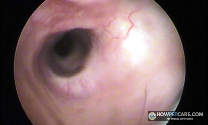

The most common dog ear problems are yeast overgrowth, bacterial infections, ear mites, allergic inflammation, foreign bodies, wax buildup, and aural hematomas. Most involve otitis externa—inflammation of the outer ear canal—usually triggered by underlying causes such as allergies, trapped moisture, or ear anatomy.
The most common dog ear problems are yeast overgrowth, bacterial infections, ear mites, allergic inflammation, foreign bodies, ear wax buildup, and aural hematomas. Most involve otitis externa—inflammation of the external ear canal—which is typically triggered by underlying factors such as allergies, trapped moisture, or ear anatomy.
Otitis externa is the most frequent ear disorder diagnosed in veterinary practices. It inflames the delicate epithelial lining of the dog’s ear canal. Unlike a human ear canal, which runs almost horizontally into the head, a dog’s ear canal has a distinct L-shaped turn from vertical to horizontal. This structure protects the delicate inner structures, but it also limits natural drainage, trapping moisture, cellular debris, wax, and opportunistic microbes. When heat and humidity build up inside the bend, normal microflora can multiply rapidly and cause painful infections.
Repeated scratching or ear flapping leaves owners wondering what’s causing the discomfort, whether it needs a clinic visit, and what that visit might cost. The answers depend on where the problem is in the ear, what allowed it to develop, and whether there are signs that need prompt veterinary attention.
Outer, middle, and inner ear infections: what separates them
Veterinarians categorize canine ear infections into three types based on how deep they occur:
- Otitis Externa (Outer Ear): Inflammation of the external ear canal, from the outer ear flap (pinna) down to the tympanic membrane (eardrum). This is the most common form of ear infection seen in primary veterinary care. Signs generally include redness, visible discharge, persistent head shaking, and an unpleasant odor.
- Otitis Media (Middle Ear): Infection of the tympanic cavity, air-filled bulla, and auditory ossicles just behind the eardrum. It most commonly develops when a severe, untreated, or chronic outer ear infection tears through the eardrum and moves inward. Over 50% of dogs with chronic, recurrent otitis externa have concurrent, often undiagnosed otitis media.
- Otitis Interna (Inner Ear): Infection of the cochlea and vestibular apparatus, which are responsible for hearing and balance. This is a serious, advanced condition. At this depth, inflammation can cause pronounced neurological signs, including involuntary eye flicking (nystagmus), a dramatic head tilt, walking in circles, nausea, and profound loss of balance.
Problems that cause infections vs. infections themselves
Bacterial and yeast overgrowths are rarely standalone diseases. They’re usually secondary opportunists that take advantage of a breakdown in the ear canal’s local defenses. Healthy canine ear canals naturally host small populations of bacteria (Staphylococcus, Pseudomonas) and yeast (Malassezia). These microbes remain harmless until a primary trigger disrupts the canal’s self-cleaning mechanism or causes mucosal inflammation.
Primary triggers include environmental allergies (atopy), food hypersensitivities, water trapped after swimming, excessive hair growth, systemic endocrine diseases like hypothyroidism, and migrating foreign matter like grass awns. Once the skin barrier breaks down, secondary microorganisms can flourish.
An aural hematoma, by contrast, is a secondary mechanical complication rather than a microbial infection. It forms when violent, repeated head shaking or aggressive scratching breaks fragile capillary beds between the ear cartilage and skin. Blood then pools in a tense, fluid-filled pocket across the ear flap. Treating the hematoma without diagnosing and resolving the underlying infection that caused the shaking will almost certainly lead to recurrence.
Signs of Ear Problems in Dogs: What Scratching, Head Shaking, and Odor Mean
Dogs can show physical signs of ear distress long before an infection causes severe structural damage. Since they can’t tell us where it hurts, owners need to watch for changes in behavior, appearance, and smell.
Behavior you can see
The earliest warning signs are almost always behavioral. Repeated actions like these can indicate irritation or pain:
- Vigorous Head Shaking: Dogs may shake their heads violently to dislodge a perceived blockage or relieve intense itching deep in the horizontal canal.
- Frequent Pawing or Scratching: Persistent kicking or scratching at the base of the ear with a hind paw, sometimes accompanied by groaning or whining.
- Rubbing Against Furniture or Carpet: Pressing the side of the face into rugs, sofas, or grass to reach an itch.
- Guarding and Sensitivity: Flinching, ducking away, growling, or yelping when someone reaches toward the head or touches the collar.
- Depression or Lethargy: Reduced appetite, reluctance to play, or more time spent sleeping because of a persistent, low-grade throb or acute pain.
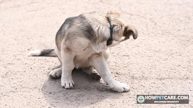
Constant scratching or shaking can quickly cause physical trauma. The force of the ear flap whipping against the skull can break internal blood vessels within hours, turning an itchy ear into a painful hematoma that may be expensive to repair surgically.
What you can smell and see inside the ear flap
A healthy canine ear canal is pale pink, dry, odorless, and free of visible buildup. Gently lifting your dog’s ear flap (the pinna) to inspect the opening can reveal useful clues:
- Visual Appearance: Redness (erythema), swelling that narrows the canal opening, raw scratches, crusting, or scaly skin along the inner flap.
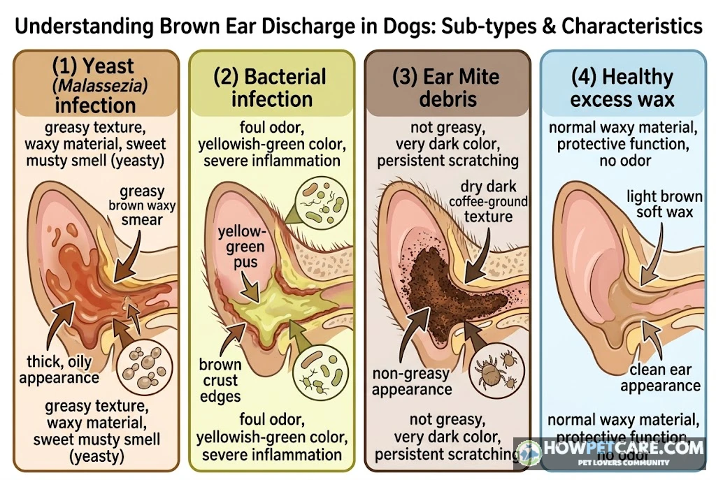
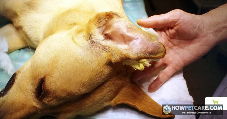
- Discharge Characteristics: Discharge varies in color and texture. You might see dry, crumbly black-brown specks resembling coffee grounds; moist, greasy golden-brown wax; or thick, purulent yellow-green pus.
- Odor Profiles: Yeast infections characteristically produce a pungent, musty, sweet smell often compared to stale beer, sourdough bread, or corn chips. Advanced bacterial infections, by contrast, can produce a foul, putrid, or rotting odor that may be noticeable several feet away.
Color and smell offer hints, but they don’t confirm a diagnosis. Dark brown discharge can come from yeast overgrowth, mixed bacterial populations, or microscopic mites. Laboratory testing is needed to know what’s growing.
Signs that point deeper than the outer ear
Signs beyond local itching and minor odor may mean an infection has breached the tympanic membrane or affected neurological pathways in the middle and inner ear. Watch for these red flags:
- A persistent, abnormal head tilt toward the affected side
- Loss of motor coordination, drunken stumbling, or leaning against walls
- Compulsive circling in one direction
- Rapid, involuntary eye movements back and forth or up and down (nystagmus)
- Drooping of the upper eyelid, lip, or ear on one side of the face, signaling facial nerve paralysis
- Sudden or progressive loss of hearing
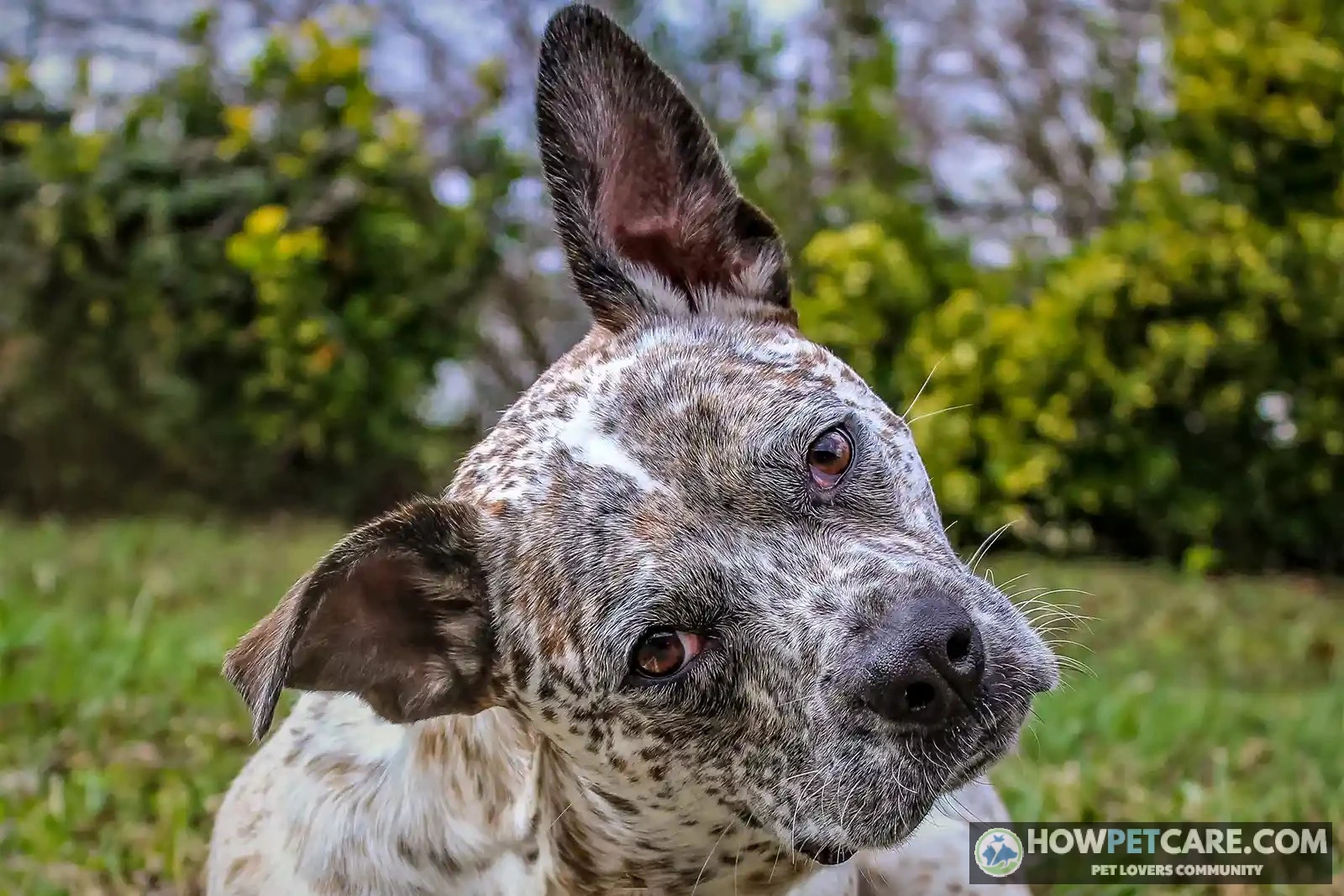
Signs in only one ear (unilateral) often point to a local physical trigger, such as a trapped foreign seed, a localized polyp, or an isolated anatomical defect. Signs in both ears at the same time (bilateral) strongly suggest an underlying systemic issue, most commonly food or environmental allergies.
Ear Mites vs. Yeast vs. Bacterial Infection vs. Foreign Body: How to Tell Them Apart
Owners naturally want to know what’s happening inside a painful ear. The visible signs overlap, but mites, yeast, bacteria, foreign objects, and hematomas each have some distinguishing characteristics.
Side-by-side symptom comparison
The table compares observable signs, risk factors, and the methods a vet uses to confirm these conditions.
| Condition | Discharge and appearance | Odor | Itch vs. pain | One ear or both | Who is most at risk or typical trigger | What the vet will confirm it with |
|---|---|---|---|---|---|---|
| Ear mites (Otodectes cynotis) | Dry, dark debris that looks like coffee grounds | Mild or musty | Intense itch, mild to moderate pain | Often both, can be one | Puppies, dogs living with cats or other pets; contagious between animals | Otoscope or microscope exam of a swab — mites are directly visible |
| Yeast infection (Malassezia) | Waxy brown or yellowish discharge, greasy-looking skin | Musty, sweet, or "corn chip" smell | Itch-driven, moderate pain | Often both | Dogs with allergies, floppy ears, or recent swimming/baths | Ear cytology (swab under microscope) showing yeast organisms |
| Bacterial infection | Yellow, green, or pus-like discharge; ear may be swollen and hot | Foul or strong smell | Pain-driven, sensitive to touch | Often one, can be both | Follows moisture, swimming, or untreated inflammation | Cytology showing bacteria; culture if severe or recurrent |
| Foreign body (grass awn, seed) | Little discharge; sometimes blood-tinged | Usually none | Sudden pain; violent head shaking or tilting | Almost always one ear | Sudden onset minutes to hours after running through grass or fields | Otoscope exam, sometimes under sedation — do not probe at home |
| Ear hematoma | No discharge; soft, puffy swelling of the ear flap itself | None | Tender rather than itchy | Usually one | After intense scratching or head shaking from another ear problem | Visual and touch exam; needle aspiration confirms blood-filled swelling |
Why these problems overlap and why cytology settles it
You can’t reliably distinguish a yeast infection from a bacterial infection by looking at discharge. Golden-brown wax often correlates with Malassezia yeast, and yellow-green fluid often points to bacteria like Pseudomonas, but mixed infections are common. A dog can have an overgrowth of yeast and rod-shaped bacteria at the same time.
Intense inflammation can also look the same whether it’s triggered by an allergy flare-up, a trapped tick, or microscopic mites. That overlap makes veterinary ear cytology essential.
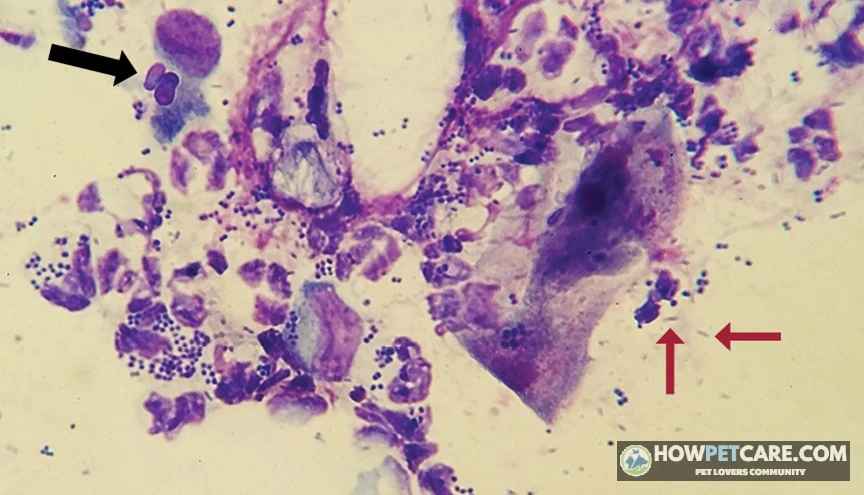
During cytology, a clinician gently inserts a sterile swab into the horizontal ear canal, rolls the collected exudate onto a glass slide, heat-fixes it, and applies specialized stains (such as Diff-Quik). Under high-power magnification, the veterinarian counts and identifies what is present: peanut-shaped budding yeast cells, round cocci bacteria, rod-shaped bacilli, inflammatory white blood cells, or live mites. Without this step, prescribing medication is guesswork that risks treatment failure and antimicrobial resistance.
Ear wax buildup, allergies, and hematoma: the look-alikes
Several non-infectious conditions can mimic or precede an active infection. Normal ear wax (cerumen) can build up heavily in dogs with overactive ceruminous glands, producing a thick, light-brown buildup without redness, pain, or odor. That’s a hygiene issue rather than an active infection.
In its early stages, allergic otitis presents as bright red, hot, itchy ear canals with minimal discharge. A dog may scratch relentlessly, leading an owner to suspect ear mites, when the cause is mucosal inflammation driven by systemic atopy or food proteins. If left unmanaged, that inflammation commonly progresses to a secondary yeast or bacterial infection.
An aural hematoma appears as a fluid-filled, fluctuant "pillow" swelling on the pinna. Owners sometimes mistake it for a giant insect bite, an abscess, or a tumor. It’s a pocket of blood from broken cartilage capillaries, caused by head shaking associated with an underlying, often overlooked infection.
Foreign bodies pose another hazard. Migrating plant seeds, particularly barbed foxtails or grass awns, can lodge deep inside the vertical canal during outdoor runs. They cause immediate, agonizing pain, and a dog may suddenly drop its head to one side, cry out, and shake uncontrollably. Foxtails have backward-pointing barbs, so each head shake drives the sharp seed deeper toward the eardrum, risking a traumatic tympanic rupture. Probing the canal with tweezers at home is dangerous and can push the awn through the delicate membrane.
 You might be wonderingDog breathing problemsDog Breathing Problems: Emergency or Not? A Triage GuideRead the answer
Monitor at Home, Book a Vet Visit, or Go Now: A Dog Ear Triage Guide
Not every ear scratch calls for an emergency hospital trip. But waiting too long to address a developing infection allows inflammation to change the structure of the ear canal. Chronic swelling thickens the canal walls (stenosis), narrowing the lumen and trapping more exudate. A problem that was easy to manage can become a stubborn, deep-seated condition.
Triage by symptom: what each tier looks like
Use this guide to choose a response and timeframe based on your dog’s current signs.
| Urgency tier | What you’re seeing | What to do | Timeframe | What not to do |
|---|---|---|---|---|
| Monitor at home | Occasional scratching; mild wax buildup; no odor, redness, swelling, or discharge; dog seems comfortable | Check the ear daily; keep it dry; note any change in a phone note or photo | 48–72 hours — if signs persist or worsen, move to "book a visit" | Don’t put any drops or cleaners in "just in case"; don’t clean more than the vet recommended |
| Book a vet visit | Noticeable odor; any discharge; persistent scratching or head shaking; redness; yelping or pulling away when the ear is touched; a repeat of a past infection; puppy or senior dog with symptoms | Call the clinic, describe the symptoms and how long they’ve lasted, and ask for a cost estimate | Within 1–2 days | Don’t wait weeks hoping it clears; don’t try leftover medication from a previous infection |
| Go now (same day) | Head tilt; loss of balance or circling; flicking eye movements; severe pain; facial droop; rapidly swelling ear flap (suspected hematoma); suspected foreign body; bleeding | Go to your vet or an emergency clinic the same day | Same day — hours matter for inner-ear signs and hematomas | Don’t probe inside the ear; don’t clean it; don’t give any medication before the exam |
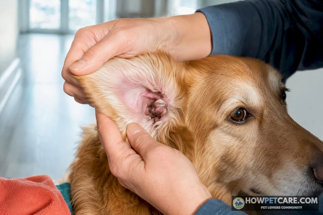
Any ear with visible discharge, redness, or heat needs a veterinary examination. The right treatment depends on identifying the microbial population and confirming that the eardrum is intact—neither can be determined at home.
What to note and photograph before you call the clinic
Before calling your veterinary provider, take two minutes to gather information that can help the triage team decide how quickly your dog needs an appointment:
- Duration and Progression: Note exactly when the scratching, shaking, or odor began. Has it steadily worsened, or did it appear out of nowhere within minutes, suggesting a foxtail or foreign body?
- Clear Visual Documentation: In a well-lit space, gently lift the ear flap and take a clear, in-focus photograph of the canal opening and inner pinna. It provides a baseline for tracking whether redness or swelling is increasing.
- Recent Environmental Context: Note whether your dog has recently been swimming in lakes or pools, groomed, bathed, or taken on field hikes through tall dry grasses within the past week.
- Current Diet and Medication History: Write down your dog’s primary food, recent treat changes, and any preventative flea/tick/mite medications. Include any past ear infection, which ear was affected, and what medication was used.
What a Vet Visit Involves, What It Costs, and How Long Recovery Takes
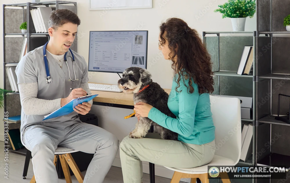
Knowing what to expect can make a veterinary appointment easier to plan and budget for. An ear examination follows several steps to identify the problem and target treatment while protecting delicate hearing structures.
How vets diagnose: otoscope, cytology, and when to go further
A comprehensive canine ear examination generally follows this sequence:
- Clinical History & Physical Assessment: The clinician reviews your dog’s symptoms, general health, lifestyle, and dermatological history.
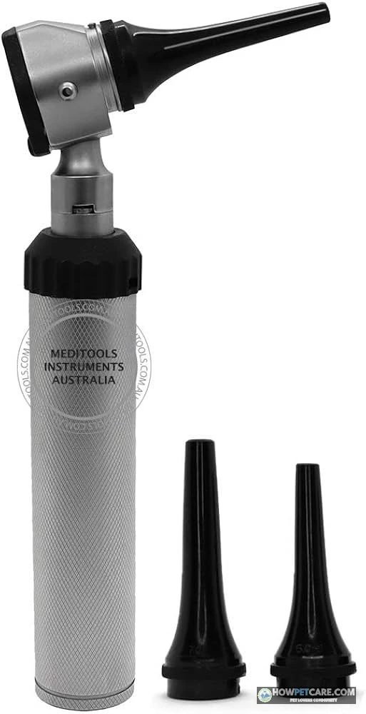
- Otoscopic Examination: Using an illuminated handheld otoscope, the vet gently guides a speculum cone down the vertical canal, past the anatomical bend, and into view of the horizontal canal. They check for tissue swelling, ulcerations, foreign material, and whether the translucent tympanic membrane (eardrum) is intact, cloudy, bulging, or torn.
- Cytological Staining: Swabs from the canal are stained and examined under a microscope to identify and quantify specific bacteria and yeast populations.
- Advanced Diagnostics: If thick exudate fills the canal or the ear is too painful to examine safely, the vet may recommend mild sedation for a thorough flush. Chronic, non-responsive, or recurrent infections may require bacterial culture and sensitivity testing to identify targeted antibiotics. Digital radiographs or CT scans can assess the deeper middle ear and bony tympanic bulla.
Treatment options by condition
Once the cause is confirmed, your veterinarian will prescribe targeted treatment:
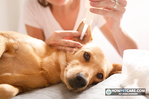
- Topical Combination Drops: Most uncomplicated outer ear infections respond to prescription topical medications such as Otomax that combine an antifungal (e.g., miconazole, clotrimazole, posaconazole), a broad-spectrum antibiotic (e.g., gentamicin, marbofloxacin, florfenicol), and a corticosteroid (e.g., dexamethasone, mometasone) to quickly relieve pain and swelling.
- Long-Acting In-Clinic Gels: For dogs that resist daily treatment at home, clinics often offer single-dose, heat-activated gel drops. The veterinary team applies them after cleaning, and the medication stays in the canal, steadily releasing active ingredients over 10 to 30 days.
- Systemic Medications: Deep-seated infections, severe otitis media, or infections with heavily ulcerated, thickened canals often require oral antibiotics, oral antifungals, or short tapering courses of oral anti-inflammatories (such as prednisone).
- Parasiticides: Ear mite infestations (Otodectes cynotis) are treated with prescription modern isoxazoline-class spot-on or chewable parasiticides, along with thorough cleaning to remove irritating mite waste.
- Hematoma Interventions: Aural hematomas require physical relief. Options include needle aspiration with steroid infusion, placement of an active silicone drain, or surgical repair under general anesthesia. In surgery, the flap is incised, drained, and sutured with staggered mattress patterns to prevent fluid from accumulating again.
Cost ranges and what drives them
Veterinary fees vary across the United States with local cost of living, the choice of general practice or emergency facility, and the dog’s size and temperament. The figures below reflect typical 2026 price ranges across the United States—always request a written estimate from your clinic before booking.
| Stage or condition | What it involves | Approximate cost range | Typical recovery time | Factors that raise the cost |
|---|---|---|---|---|
| Vet exam + ear cytology | History, otoscope exam of canal and eardrum, microscope exam of a swab | $75–$200 combined | Same visit | Emergency or after-hours clinics; specialist consults |
| Uncomplicated outer ear infection | Topical ear drops (antibiotic/antifungal/anti-inflammatory), take-home cleaner | $25–$100 for medication on top of the exam | 1–2 weeks | Severe infections needing oral medication add $30–$80 |
| Ear flush under sedation | Deep cleaning of a painful or blocked canal | $100–$300 | Cleaning itself is same-day; underlying infection still needs 1–4 weeks of treatment | Anesthesia monitoring, culture testing |
| Ear mites | Prescription mite treatment (often a topical spot-on), treatment of in-contact pets | $40–$150 | 2–4 weeks including follow-up | Treating multiple pets in the household |
| Hematoma repair | Drainage or surgical repair of the ear flap | $300–$800 | 2–3 weeks for flap healing | Size of hematoma, sedation needs, concurrent ear infection treatment |
| Middle or inner ear infection | Oral medication, possible imaging (X-ray or CT), deep flush | $200–$1,500+ depending on imaging | 3–6 weeks | CT imaging and specialist referral |
| TECA (total ear canal ablation) | Surgical removal of a permanently diseased ear canal | $3,000–$5,000+ | 3–4 weeks surgical recovery | Specialist surgeon fees, bilateral surgery |
Don’t treat the follow-up appointment as optional. Factor a recheck visit ($40–$80) into your initial budget. An ear can look cured on the outside while active, resistant microbes remain deep near the eardrum. Stopping treatment early without a clear cytological recheck is the leading cause of immediate relapse.
Realistic recovery timeline by condition
Healing times depend on how deep the problem is and how quickly treatment begins:
- Uncomplicated Otitis Externa: Typically resolves in 1 to 2 weeks with appropriate topical formulations.
- Standard Moderate Bacterial or Fungal Infections: Usually require 2 to 4 weeks of consistent medical management to clear both the clinical signs and microscopic organisms.
- Deep Middle or Inner Ear Disease (Otitis Media/Interna): Requires sustained systemic antimicrobial and anti-inflammatory therapy for 3 to 6 weeks, with serial re-evaluations.
- Minor Eardrum Perforations: Small tears in the tympanic membrane usually heal on their own in 2 to 3 weeks, provided the canal is kept free of toxic substances and ongoing infection is controlled.
- Aural Hematoma Healing: Surgical incisions and ear cartilage require 2 to 3 weeks to heal securely and allow suture removal.
How to Clean a Dog’s Ears Safely: Eardrum Check, Steps, and What Never to Use
Routine cleaning at home can help prevent problems in dogs prone to wax buildup or those that swim frequently. But putting cleaner into an infected, ulcerated, or structurally damaged ear can cause severe pain and risk lasting neurological harm.
Before you start: is it safe to put anything in the ear?
Before using an ear cleaning solution, confirm that the ear is safe to clean. Never pour liquid or drops into your dog’s ear if you see:
- Severe pain or intense distress when the ear is lightly touched
- A canal that appears completely closed, severely narrowed, or swollen shut
- Oozing blood, thin reddish serum, or thick creamy green pus
- A persistent head tilt, balance problems, or eye flickering
These signs often point to a ruptured eardrum or an active middle ear infection. The eardrum is a delicate barrier that protects the middle and inner ear. If it’s perforated, common veterinary ear cleaning ingredients (including certain detergents, drying agents, and aminoglycoside antibiotics) can enter the middle ear chamber and damage the auditory ossicles and vestibular nerve. That can cause sudden deafness, persistent loss of balance, or facial nerve paralysis.
You can’t see the eardrum at home. Always have your veterinarian examine an irritated ear with an otoscope and confirm the eardrum is intact before applying at-home cleaners.
Step-by-step ear cleaning
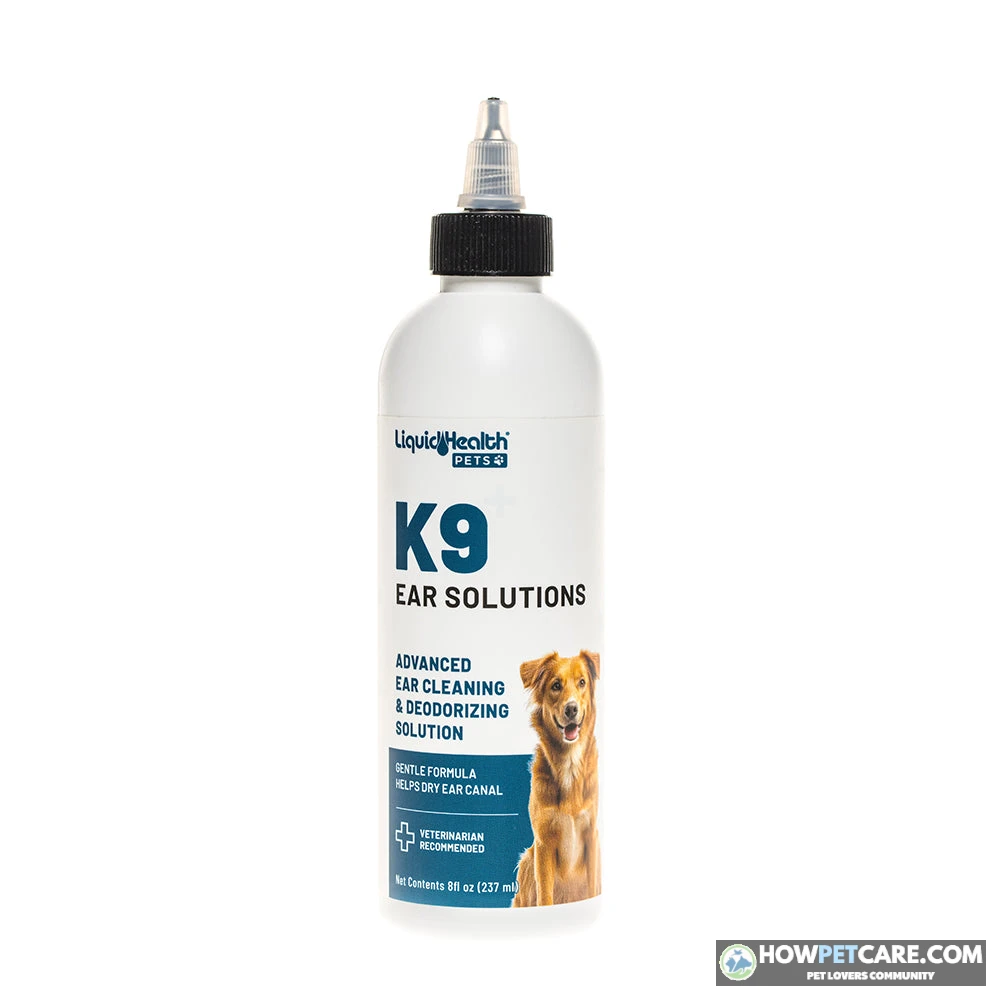
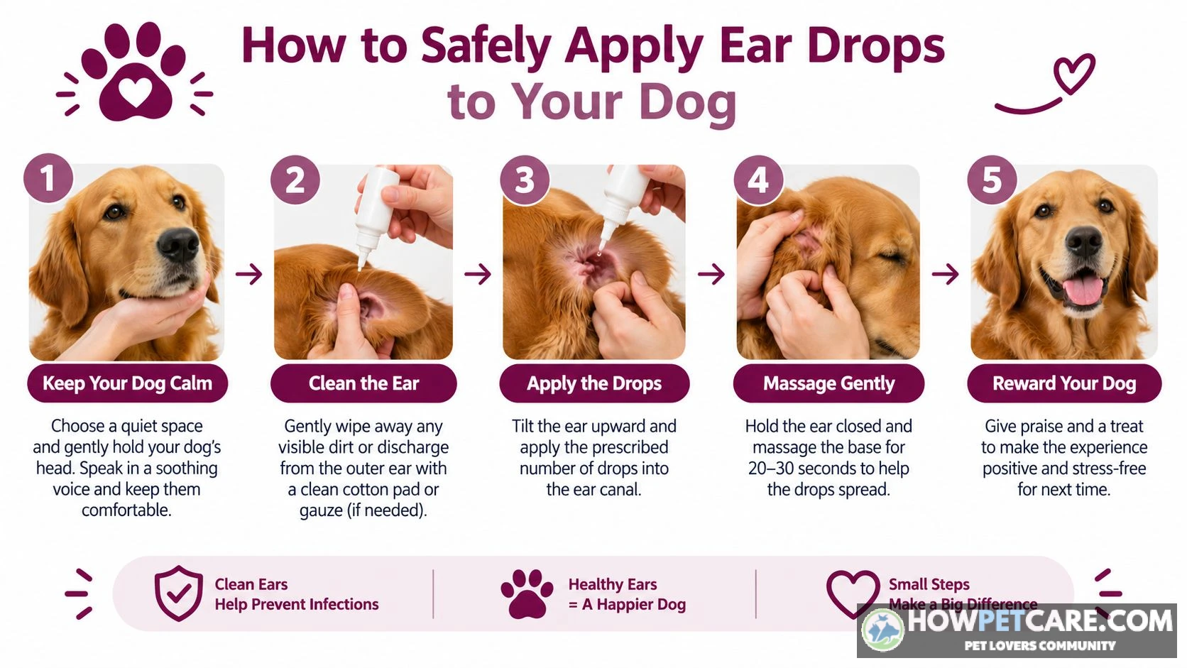
For a healthy, non-painful ear cleared for regular maintenance, use a quality, vet-formulated canine ear cleaning solution. Follow this five-step technique:
-
Position the ear and prep your supplies
Gently grasp the tip of the ear flap (pinna) and lift it upward and slightly back to straighten the L-shaped ear canal. Keep the cleaning solution and several pieces of soft medical gauze or cotton balls within easy reach.
-
Fill the canal with solution
Place the nozzle of the cleaning solution bottle at the entrance of the open canal and squeeze generously until the canal is comfortably filled. Don’t force the plastic bottle tip deep into the ear.
-
Massage the base of the ear
Hold the ear flap upright and use your thumb and forefinger to massage the cartilaginous base of the ear just below the opening for 20 to 30 seconds. You should hear a wet squelching sound. The gentle movement of fluid breaks up waxy plugs and dislodges debris clinging to the horizontal canal walls.
-
Step back and let the dog shake
Release the ear and step back. Your dog will naturally shake its head vigorously, lifting loosened debris, wax, and excess liquid from the horizontal canal into the outer ear flap where you can reach it.
-
Wipe away loosened debris
Wrap a clean piece of cotton gauze around your finger and gently wipe loosened wax, exudate, and remaining cleaner from the inner ear flap and visible canal opening. Clean only as far as your finger naturally reaches—never force materials deeper.
Home remedies to avoid and why
Online forums and social media often suggest DIY solutions for ear problems. These unverified mixtures and tools can cause serious complications:
- Cotton Swabs (Q-tips): Never insert cotton swabs into your dog’s ear canal. The canal turns horizontally at a 90-degree angle, and a rigid swab acts like a piston, packing wax, exudate, and microorganisms against the eardrum. It can also puncture the tympanic membrane if the dog jerks unexpectedly.
- Hydrogen Peroxide: Peroxide bubbles aggressively when exposed to biological enzymes, causing mechanical irritation. It adds unwanted moisture and breaks down newly forming epithelial cells, worsening tissue damage.
- Rubbing Alcohol (Isopropyl): Alcohol is intensely painful on raw, scratched, or inflamed skin. It rapidly dehydrates delicate tissues, causing stinging and defensive aggression.
- Vinegar Solutions (Acetic Acid): Undiluted or improperly balanced vinegar burns raw, inflamed ear tissue. Although acetic acid has mild antibacterial qualities in balanced laboratory formulations, homemade vinegar mixes disrupt the canal’s natural pH and cause painful chemical irritation.
You can’t safely or successfully treat an established microbial dog ear infection at home without veterinary intervention. Home remedies don’t have the targeted potency to eliminate deep bacterial colonies or fungal overgrowths. Relying on them delays proper care and allows infections to spread deeper into the middle ear.
For routine home care, make a habit of a quick, weekly visual ear check. Keep ears dry after every swimming session, and ask your veterinarian about an appropriate cleaning frequency. Over-cleaning an otherwise healthy ear can irritate it and disrupt its natural defenses.
Dog Breeds Most Prone to Ear Problems and the Anatomy Behind It
Any dog can develop an ear infection, but some breeds are structurally predisposed to recurrent ear disease. Anatomy, genetics, and lifestyle all influence how easily an ear canal traps moisture and debris.
Why floppy ears, hair, and narrow canals raise risk
The canine ear canal relies on airflow to evaporate normal moisture and maintain an environment that’s inhospitable to yeast and bacteria. Several physical features can interfere with that defense:
- Pendulous Pinnae (Floppy Ears): Heavy, hanging ear flaps cover the canal opening and block airflow. This creates a dark, warm, humid microclimate that encourages rapid microbial growth.
- Hyper-Trichoic Canals (Hairy Canals): Dense hair inside the ear canal catches shedding epithelial cells, traps cerumen, and holds moisture against the skin.
- Stenotic or Folded Canals: Highly folded or narrowed ear canals restrict air movement and prevent wax and debris from migrating naturally outward.
- Atopic Tendencies: Genetic predispositions to environmental allergies (atopic dermatitis) cause chronic inflammation of the skin barrier, including the ear canal lining.
Breed groups at higher risk
The table outlines breeds more susceptible to ear issues, the mechanisms involved, and practical prevention priorities.
| Risk mechanism | Higher-risk breeds | What’s driving it | Prevention focus |
|---|---|---|---|
| Floppy, heavy ear flaps | Cocker Spaniels, Basset Hounds, Beagles, Labrador and Golden Retrievers | The flap blocks airflow, trapping heat and moisture — the environment yeast and bacteria thrive in | Weekly ear checks; lift the flap to air the ear; dry thoroughly after baths and swimming |
| Hairy ear canals | Poodles, Schnauzers, Bichon Frises | Hair inside the canal traps wax and debris and holds moisture against the skin | Vet-guided hair management — ask before plucking, since it can irritate some ears |
| Narrow or heavily folded canals | Shar-Peis, Bulldogs | A tight, folded canal ventilates poorly, so inflammation has nowhere to drain | Regular checks; early vet visits at the first sign, since these ears escalate faster |
| Inherited allergy tendency | Labradors, Golden Retrievers, French Bulldogs, West Highland White Terriers | Allergies inflame the canal lining; the infection is a symptom, the allergy is the cause | Managing the underlying allergy with your vet — cleaning alone won’t prevent flare-ups |
| Frequent swimming | Any water-loving breed, especially retrievers and spaniels | Water left in the canal softens the skin and invites overgrowth | Dry the outer ear after every swim; ask your vet about a drying routine |
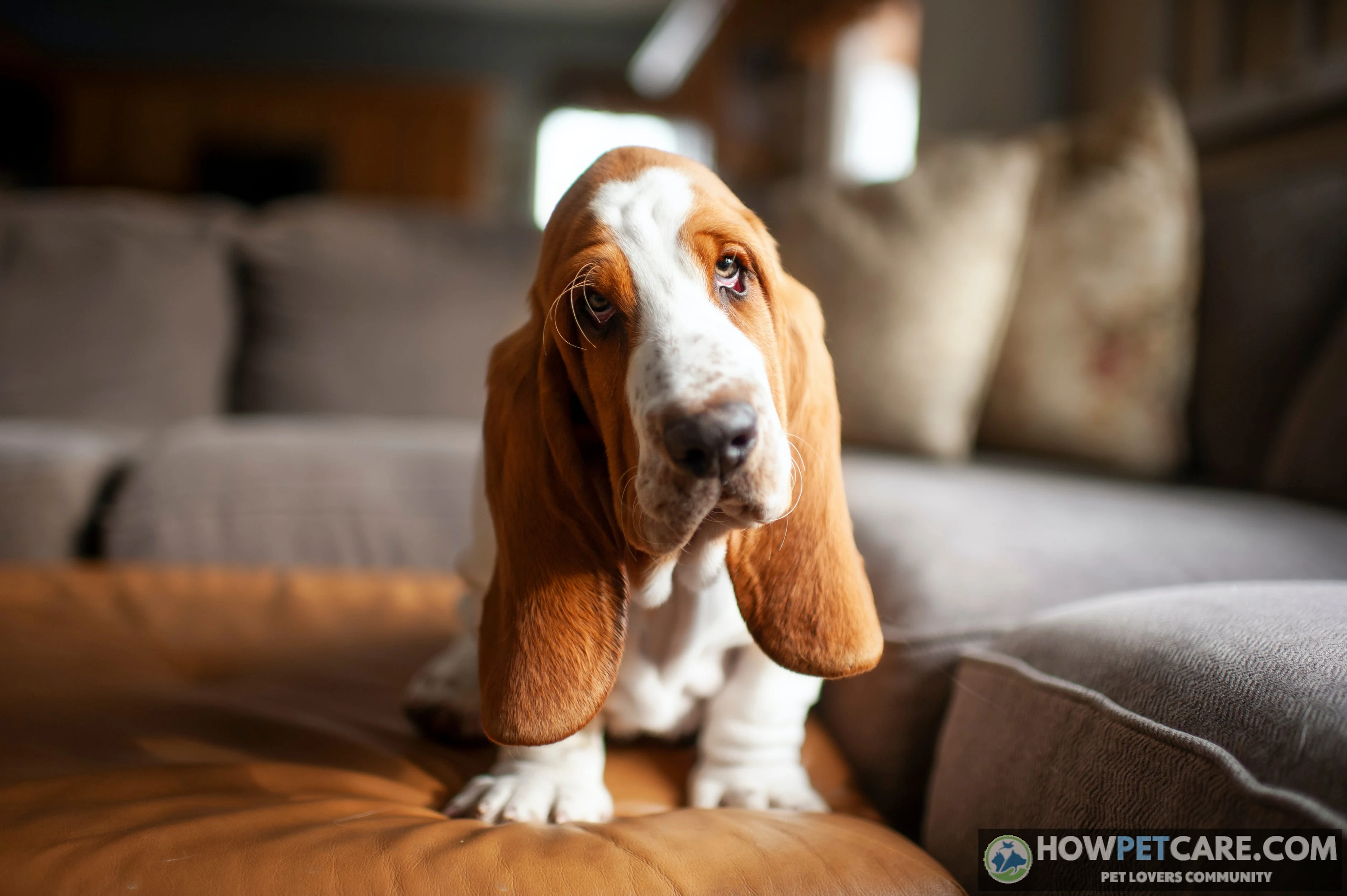
Breed predisposition is a risk factor, not a guarantee. Upright-eared dogs like German Shepherds and Huskies can also develop severe infections if they have allergies or foreign bodies. Prevention works best when it addresses the underlying mechanism—minimizing moisture, monitoring hair accumulation, and managing allergies—rather than relying on frequent ear cleaning alone.
Why Dog Ear Infections Keep Coming Back: Allergy Workups, Hidden Middle-Ear Disease, and Long-Term Management
An ear infection that clears up only to return a few weeks later can be frustrating. Recurrence is rarely due to failure of the original antibiotic. Instead, it almost always means the primary underlying driver wasn’t resolved or that infection remains hidden deeper in the ear.
Why the same infection returns
Bacterial and yeast overgrowths are secondary symptoms. Clearing the active microbial population with medication gives the dog temporary relief. But if the underlying trigger continues, the ear canal lining becomes inflamed again, creating conditions for microbes to recolonize.
Common drivers of chronic relapses include:
- Underlying Allergic Disease: Atopic dermatitis (reactions to airborne pollens, grasses, or house dust mites) and food allergies are by far the leading causes of recurrent canine ear problems. Recurrent ear inflammation is often the first and sometimes the only clinical symptom of food or environmental allergies.
- Undiagnosed Middle Ear Disease (Otitis Media): In over 50% of chronic, recurring otitis cases, bacteria have breached the eardrum and established a persistent infection inside the middle ear bulla. Drops placed in the outer ear can’t clear infection behind an intact or partially healed eardrum. After outer treatment ends, bacteria from the middle ear can migrate outward and re-infect the canal.
- Endocrine Imbalances: Unmanaged systemic conditions, particularly hypothyroidism or Cushing’s disease, alter normal sebum production and suppress cutaneous immune defenses, leaving ears vulnerable to opportunistic overgrowths.
What an allergy workup and elimination diet look like
When a dog has two or three ear infections within a single year, your veterinarian will likely recommend moving beyond treatment of each infection to a formal allergy workup:
- Eliminating Ectoparasites & Secondary Infections: First, the vet confirms that active microbial overgrowths have cleared and rules out parasites like mites or fleas.
- Strict Dietary Elimination Trial: To diagnose or rule out food hypersensitivities, the dog undergoes a dedicated 8 to 12-week dietary trial. The dog eats exclusively a veterinary-prescribed hydrolyzed protein diet (where proteins are broken down so small the immune system cannot detect them) or a novel single-protein diet (such as venison or rabbit) that the dog has never eaten before. During these 8 to 12 weeks, the dog must not receive standard treats, table scraps, flavored supplements, or chew toys. If ear inflammation resolves completely during the trial, food components are reintroduced one by one to confirm specific food allergies.
- Environmental Allergy Management (Atopy): If food allergies are ruled out, environmental atopy is the primary suspect. Long-term options include targeted allergy testing, allergen-specific immunotherapy (hyposensitization injections or drops), or modern immunomodulating medications (such as Apoquel or Cytopoint) that suppress the allergic itch reflex.

When chronic ear disease needs a specialist or surgery
In severe, long-standing chronic otitis, repeated cycles of uncontrolled inflammation permanently change the ear canal’s anatomy. The walls thicken with heavy scar tissue (hyperplasia), develop obstructive, calcified folds, and narrow into tight crevices that drops can no longer reach and cleaning can’t effectively clear. Eventually, flexible cartilage can undergo dystrophic calcification, turning the soft ear canal into rigid, mineralized bone.
When an ear reaches this irreversible, painful, end-stage condition—or when middle ear disease doesn’t respond to deep flushes and medical therapy—a board-certified veterinary surgeon may recommend a Total Ear Canal Ablation with Lateral Bulla Osteotomy (TECA-LBO).
TECA is a salvage procedure that permanently removes the entire diseased external ear canal and clears infected bone from the middle ear bulla. Although hearing in that ear is significantly reduced, the dog’s inner ear can often still detect vibrations. Removing the chronically infected, painful tissue relieves the persistent discomfort of end-stage ear disease.
TECA is an advanced, last-resort surgery reserved for end-stage ears. The vast majority of chronic ear cases can be managed without surgery through accurate cytology, structured allergy workups, and a consistent veterinary maintenance routine.
Your Dog Ear Problem Action Plan: A Step-by-Step Checklist
Use this checklist to move from noticing symptoms to getting your dog relief:
- Check for Emergency Red Flags: Look at your dog’s eyes, head posture, and movement. If you notice a head tilt, loss of balance, circling, abnormal eye flicking, or a rapidly ballooning ear flap, skip at-home observation and contact your vet or an emergency clinic immediately.
- Evaluate Observable Symptoms: Compare the discharge color, smell, and your dog’s behavior with the symptom comparison table to narrow down what might be happening.
- Determine Your Triage Tier: Decide whether your dog’s signs call for 48 to 72 hours of monitoring, a standard veterinary appointment within 1 to 2 days, or same-day urgent care.
- Close the Eardrum Gate: Don’t put leftover medications, over-the-counter drops, cleaners, or home remedies into the ear until a veterinarian has examined the eardrum with an otoscope.
- Prepare for the Appointment: Note when symptoms started, take a well-lit photo of the ear, write down your dog’s current diet and medications, and ask for a diagnostic estimate that includes exam and cytology fees.
- Complete the Prescribed Course: Give every dose exactly as directed by your vet, even if the ear looks completely healed after a few days. Attend the scheduled recheck to confirm the infection has cleared microscopically.
- Address the Primary Cause: If this is your dog’s second or third ear issue, talk with your vet about investigating underlying causes, including an 8 to 12-week elimination diet trial or environmental allergy testing.
For more detail, see our related guides on dog ear discharge colors, chronic ear odor, persistent head shaking, safe ear cleaning solutions, inner-ear growths and polyps, and surgical options for aural hematomas.
 You might be wonderingDog peeing bloodDog Peeing Blood: Emergency Signs, Causes, and Next StepsRead the answer
FAQ
Can a dog’s ear problem go away on its own?
Mild wax buildup or minor irritation sometimes resolves, but infections, mites, and foreign bodies rarely do and tend to get worse. If your dog has odor, discharge, or ongoing scratching, have a vet examine the ear.
Are dog ear infections contagious to other pets?
Ear mites spread easily between dogs, cats, and other pets, so all pets in the household usually need to be checked. Yeast and bacterial infections generally aren’t contagious, because they develop from an underlying problem in the individual dog’s ear.
Will an ear hematoma heal without treatment?
A hematoma may slowly reabsorb, but it stays painful for weeks and often leaves a scarred, crumpled ear. A vet should treat both the hematoma and the cause of the head shaking behind it.
How often should I clean my dog’s ears?
It depends on the dog. Many dogs with healthy ears need little or no routine cleaning, while floppy-eared, swimming, or allergic dogs may need regular cleaning. Ask your vet to set a schedule, because over-cleaning can irritate the ear.
Can I use human ear drops or medications on my dog?
No. Human products may be irritating or unsafe for dogs, and they can do harm if the eardrum is damaged. Use only products your vet has chosen after examining the ear.