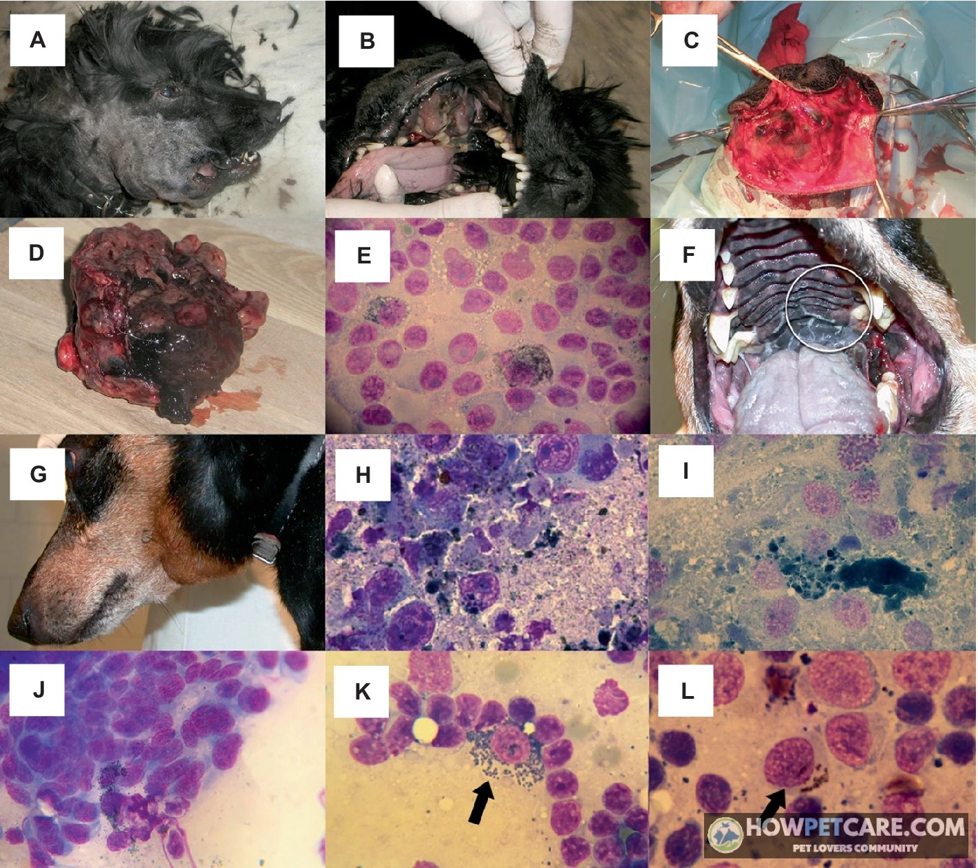

Melanoma in dogs is a tumor of pigment-producing melanocyte cells that most often appears in the mouth, on the skin, at the nail bed, or in the eye. About a third lack pigment and look pink rather than black. Only a biopsy can confirm it — oral and nail-bed forms are usually the most aggressive.
If you’ve found a dark, pink, or flesh-colored growth on your dog’s mouth, skin, toe, or eye, you may be wondering what melanoma looks like and whether the growth is an emergency. Schedule a veterinary exam promptly, and make any mouth, nail-bed, or eye growth a same-week priority. Melanoma in dogs begins in melanocytes, the cells that produce pigment. Some canine melanomas are benign, but oral and nail-bed melanomas often grow aggressively and can spread to lymph nodes or the lungs. Color alone can’t tell you whether a lump is dangerous. About one-third of canine melanomas are amelanotic, meaning they have little or no pigment and may look pink or skin-colored rather than black. Only a tissue sample, such as a fine needle aspiration or biopsy, can confirm what the growth is. Early signs include a new dark or pink lump, bleeding, odor, drooling, limping, or a sore that won’t heal. This guide covers how melanoma looks in different locations, diagnosis and staging, treatment, the limits of the Oncept melanoma vaccine, expected costs in the United States, and questions to take to your first appointment.
A new lump may be melanoma—even if it is pink: what to do first
Book a veterinary visit within days if the growth is new, growing, bleeding, ulcerated, painful, or hasn’t gone away after a week or two. Seek urgent or emergency care the same day if your dog has trouble breathing or swallowing, bleeding that won’t stop, severe pain, or a sudden loss of function, such as being unable to walk on a toe or eat normally. A small, stable lump on haired skin can usually wait one to two weeks for a routine appointment, but it still needs a check.
While you wait, photograph the mass next to a ruler or coin in good light from two angles. Repeat the photos weekly to document any growth. Write down when you first noticed it and any changes in size, color, bleeding, odor, scratching, or licking. Don’t squeeze, cut, bandage over, or apply home remedies to the lump. These actions can interfere with testing and make the situation worse.
A dark lump isn’t automatically cancer, and a pink lump isn’t automatically safe. Roughly a third of canine melanomas have little or no pigment. Location and biopsy—not color—determine seriousness. Never apply human skin creams, essential oils, or “drawing salves” to a mass before a vet examines it.
What a suspicious growth can look like by location
| Location | Possible melanoma appearance or signs | Benign or other look-alikes | When to seek care |
|---|---|---|---|
| Mouth (gums, lips, tongue, palate) | Dark OR pink/flesh-colored mass; may bleed, ulcerate, or smell foul; drooling, dropping food, reluctance to chew | Gum overgrowth (epulis/gingival hyperplasia), viral papillomas (warts), dental abscess | Book within days for any oral mass. Emergency if bleeding won’t stop or your dog can’t eat or breathe comfortably |
| Haired skin | Small, firm, dark nodule; some are pink or skin-colored (amelanotic); may grow, itch, or ulcerate | Benign melanocytoma, histiocytoma, skin tag, sebaceous cyst, attached tick | Routine appointment within 1–2 weeks for a stable small lump; sooner if it’s growing fast, bleeding, or changing color |
| Nail bed / toe | Swollen toe, deformed or missing nail, lump at the nail base, limping, constant licking, wound that won’t heal | Broken nail, nail-bed infection (paronychia), interdigital cyst | Prompt visit if nail loss plus swelling persists more than 1–2 weeks, or the toe keeps getting bigger |
| Eye / eyelid | Dark or pink mass on the lid, iris, or conjunctiva; squinting, tearing, a cloudy or enlarged eye, vision change | Meibomian (eyelid gland) tumor, conjunctival cyst, benign iris freckle | Prompt exam; urgent if the eye is painful, bulging, or your dog seems to be losing vision |
Key rule: color alone can’t confirm or rule out melanoma — only a tissue sample can.

What to record before the appointment
- The date you first noticed the growth and how it has changed.
- Whether your dog is licking, scratching, or avoiding use of the area.
- Any related changes: bad breath, drooling, dropping food, limping, squinting, or new lumps elsewhere — any of which can also be warning signs of cancer in dogs.
- All medications and supplements your dog currently takes.
How location changes the risk: mouth, skin, nail bed, and eye
Where a melanoma sits tends to be the biggest factor in how serious it is, not how dark it looks. Canine melanoma behaves differently depending on whether it arises in the mouth, on the skin, at the nail bed, or in the eye. Melanoma accounts for roughly 30 to 40 percent of oral tumors in dogs. Most mouth and nail-bed melanomas are malignant and invasive, whereas most melanomas on haired skin are benign.
Oral melanoma
Oral melanoma in dogs often behaves aggressively. It can invade the jawbone, spread to nearby lymph nodes, and metastasize to the lungs. Warning signs include a dark or pink mass on the gums, lips, tongue, or palate; bleeding from the mouth; foul breath; drooling; dropping food; and reluctance to chew or play with toys. These tumors are often advanced by the time owners notice them, so any oral mass should be examined within days. Other cancers, such as squamous cell carcinoma, can cause similar oral signs. Prognosis depends heavily on stage and whether the tumor can be removed completely, making staging and imaging especially important for this location.

Cutaneous and amelanotic melanoma
Cutaneous melanoma on haired skin is often benign; studies suggest 85 to 90 percent of these tumors are non-malignant. A changing, ulcerated, fast-growing, or unpigmented skin mass still needs testing because it can be malignant. Some growths called benign melanocytomas look like dark, firm nodules but don’t spread; only pathology can distinguish them from malignant melanoma. Dark spots aren’t the only warning sign. About one-third of canine melanomas are amelanotic, meaning they lack pigment and may appear pink or flesh-colored. These growths are easy to miss, so any new skin nodule that’s growing or changing should be sampled.

Nail-bed and ocular melanoma
Digital or nail-bed melanoma may cause a swollen toe, a deformed or missing nail, a lump at the nail base, limping, constant licking, or a sore that won’t heal. Treatment often involves amputation of the affected toe. Reported average survival after digit amputation is about 12 months, and one study cited 476 days when amputation was combined with the Oncept vaccine. Ocular melanoma varies according to which eye structure is involved. Squinting, tearing, a cloudy or enlarged eye, a visible mass on the lid or iris, or a change in vision merits prompt examination. A painful or bulging eye is more urgent.

Diagnosis and staging: what the vet checks and what Stage I–IV mean
Your veterinarian will likely measure and examine the mass, check nearby lymph nodes even if they don’t feel enlarged, and recommend a tumor sample. Fine needle aspiration uses a thin needle to withdraw cells for review under a microscope. A biopsy removes a small piece of tissue under sedation or anesthesia for histopathology, the lab report that identifies tumor type, margins, and sometimes mitotic rate (how quickly the tumor cells are dividing). Depending on the tumor’s location and your treatment goals, staging can include chest X-rays to look for spread to the lungs, abdominal ultrasound, or CT scanning.

From lump sample to pathology report
The pathologist’s report will usually say whether the growth is a benign melanocytoma or a malignant melanoma. If the mass was removed, it will also usually say whether the margins are clean. For oral tumors, the report may address invasion into bone. This information guides the treatment plan and helps determine the stage.
A stage-and-survival guide for oral melanoma
Veterinarians commonly use a size-and-spread framework for oral melanoma. It doesn’t apply to skin, nail-bed, or eye melanomas, which are assessed differently. Stage I is a tumor under 2 centimeters with no lymph-node or distant spread; Stage II is 2 to 4 centimeters with no detectable spread; Stage III is over 4 centimeters and/or spread to nearby lymph nodes; and Stage IV means distant metastasis, most often in the lungs. The reported median survival times below come from studies of dogs treated primarily with surgery. A median means half of the dogs in those studies lived less time and half lived longer; it isn’t a prediction for your dog.
| Stage | Tumor size and spread | Reported median survival | Important context |
|---|---|---|---|
| Stage I | Tumor under 2 cm; no lymph-node or distant spread | Roughly 12–18 months | Best outcomes; some dogs achieve long-term control with complete surgical removal |
| Stage II | Tumor 2–4 cm; no detectable spread | Roughly 5–8 months | Larger tumors are harder to remove with clean margins; adding radiation may improve local control |
| Stage III | Tumor over 4 cm and/or spread to nearby lymph nodes | Roughly 3–6 months | Node involvement signals more aggressive disease; combined surgery + radiation ± vaccine is often discussed |
| Stage IV | Distant metastasis (most often lungs) | Roughly 1–4 months | Treatment usually shifts toward comfort; some owners explore clinical trials |
Where studies disagree: these figures are medians pooled from older studies of dogs treated mostly with surgery. Newer studies using surgery plus radiation and the melanoma vaccine sometimes report longer survival, while untreated dogs generally fall below these numbers. Ask your vet which stage—and which treatment—the number assumes.
Published survival times reflect groups of dogs in past studies. Your dog’s outlook depends on tumor location, stage, whether the entire mass could be removed, and which treatments are used. No article can predict an individual dog’s lifespan.
Treatment options, from tumor removal to newer radiation approaches
Treatment depends on the tumor’s location and stage, whether the mass can be fully removed, and your goals for your dog’s comfort and longevity.
Surgery, radiation, and chemotherapy
Surgery is usually the first choice when complete removal is feasible. The aim is to take the tumor with wide margins—a border of healthy tissue around it—to reduce the chance of regrowth. Oral tumors may require mandibulectomy or maxillectomy, removal of part of the lower or upper jaw, while a digit tumor often calls for amputation of the affected toe. Surgery alone can cure some localized, benign skin tumors, but oral melanoma often needs additional therapy because it tends to spread.
Radiation therapy may be recommended when complete surgery is difficult, as with some oral or nail-bed tumors, or to target cancer cells remaining after surgery. Conventional radiation involves multiple sessions over several weeks, usually with anesthesia for each visit. Stereotactic radiation (SRS) delivers one to three high-precision treatments and is available at limited referral centers. It’s worth asking about if your dog is a candidate and you can travel to a specialty hospital.
Chemotherapy is sometimes discussed for canine melanoma, but evidence of a survival benefit is limited and inconsistent. Some sources describe it as largely ineffective for melanoma, while others note possible benefit in selected cases. Chemotherapy isn’t a reliable standalone cure. Rather than assuming it will help or dismissing it outright, weigh its role carefully with a veterinary oncologist.

Oncept vaccine: what the evidence can and cannot say
The Oncept melanoma vaccine is a DNA-based immunotherapy licensed for use in dogs. It helps the immune system recognize tyrosinase, a protein associated with melanoma cells. It isn’t a preventive vaccine or a standalone cure. It’s generally considered an adjunctive option for dogs with oral melanoma that has been locally controlled—often stages II and III—and is typically given after surgery or radiation. The vaccine may stimulate an immune response, but a definitive survival benefit hasn’t been proven in all studies. Whether it’s worth considering depends on your dog’s tumor type, stage, and treatment plan. Ask a veterinary oncologist rather than assuming it’s appropriate for every case.

Palliative care and newer options
When curative treatment isn’t possible or disease is advanced, palliative care focuses on pain relief, nutrition, and comfort. Radiation can sometimes be used palliatively to shrink a painful tumor. Clinical trials and referral to a veterinary oncology specialist are also options for owners who want to explore newer approaches beyond standard care.
Prognosis, progression, and quality of life
Whether melanoma is curable depends heavily on location, stage, margins, and spread. Some localized skin tumors can be controlled or removed with surgery, while oral or metastatic disease is more challenging. Without treatment, an aggressive oral or nail-bed tumor can grow locally, interfere with eating or walking, and may spread to lymph nodes or the lungs. Progression differs from dog to dog, and no one can give an exact timeline.

How to assess comfort over time
Track pain, appetite, mobility, breathing, social engagement, and good versus bad days. One bad day is only part of the picture; look at the weekly pattern.
Daily comfort log — copy this into a notebook or notes app:
Date: ______

[ ] Ate a reasonable amount of food        0 / 1 / 2
[ ] No obvious pain (whimpering, hiding,
    guarding the mouth or limb)             0 / 1 / 2
[ ] Breathing comfortably at rest          0 / 1 / 2
[ ] Moved around / walked without struggle 0 / 1 / 2
[ ] Engaged with family or toys            0 / 1 / 2
[ ] Slept through most of the night        0 / 1 / 2

Total: ___ / 12        Today was:  GOOD / BAD
Notes (bleeding, skipped meals, new swelling):
_______________________________________________
How to use it: score each item 0 (bad), 1 (okay), or 2 (good) once a day. If bad days outnumber good days across a week, or the total drops below roughly 7 and stays there, call your vet. That’s a signal to revisit the treatment plan or discuss comfort-focused care. Bring the log to appointments so decisions are based on trends, not memory.
When to revisit goals of care
End-of-life decisions should center on your dog’s suffering and ability to enjoy daily life, not on extending time at any cost. Your veterinarian can help you identify the specific signs that mean comfort-focused care or euthanasia is the kindest choice.
Talk with your veterinarian about what decline looks like for your dog’s specific tumor and when euthanasia may be the kindest option. These decisions should center on suffering and your dog’s ability to enjoy daily life. Pain relief, appetite support, and other palliative measures can often extend good days even when the cancer itself can’t be stopped.
Diagnosis and treatment costs: plan for the steps, not one quoted total
Costs vary widely by region, clinic type, tumor location, and case complexity. Melanoma treatment for dogs can range from a few hundred dollars for a simple skin lump to well over ten thousand for advanced oral cancer care. The figures below are indicative US-dollar ranges, not guarantees. Always request a written, itemized estimate before proceeding. A commonly cited overall range for canine melanoma treatment is roughly $350 to $12,000, but that span covers everything from a simple skin lump removal to advanced oral cancer care.
| Care step | What it may include | Indicative cost range | Factors that change the estimate |
|---|---|---|---|
| General exam | Office visit, oral/skin check, mass measurement | $60–$150 | Emergency vs scheduled visit; region |
| Fine needle aspiration | Needle sample of lump and/or lymph node + cytology read | $80–$250 | Sedation adds cost; sampling multiple sites costs more |
| Biopsy + histopathology | Tissue sample under anesthesia; lab report on tumor type, margins, mitotic rate | $400–$1,200 | Oral biopsies often cost more; full report with grading adds lab fees |
| Staging imaging | Chest X-rays ($150–$350), abdominal ultrasound ($300–$600), CT ($1,200–$2,800) | $300–$3,000 | How extensive the spread work-up needs to be |
| Oncology consultation | Specialist review, treatment plan | $150–$350 | University vs private specialty hospital |
| Skin mass removal | Excision of a haired-skin tumor with margins | $600–$2,000 | Size, location, whether a specialist is needed |
| Digit amputation | Removal of the affected toe | $1,000–$2,500 | Aftercare and pathology usually included |
| Jaw surgery | Mandibulectomy or maxillectomy (removal of part of the jaw) | $2,500–$6,000+ | Complexity of reconstruction, ICU stay, complications |
| Radiation — conventional | Multiple sessions over several weeks | $4,000–$7,500 | Number of sessions; anesthesia each visit |
| Radiation — stereotactic (SRS) | 1–3 high-precision treatments | $4,500–$8,500 | Only available at limited referral centers |
| Oncept vaccine | 4 doses every 2 weeks, then a booster about every 6 months | $2,500–$3,500 in year one | Ongoing booster costs; usually added after local control |
| Chemotherapy (selected cases) | Drug cost plus monitoring bloodwork | $300–$800 per treatment | Number of cycles; evidence for benefit is limited |
Scenario totals to budget around: removal of a haired-skin melanocytoma or melanoma with pathology often costs near $1,000–$2,500 all-in. An oral Stage II case with full staging, jaw surgery, and a vaccine series commonly totals $6,000–$12,000 or more. Ask every provider two questions: “Which of these tests will change what we do next?” and “What’s the palliative-only version of this plan and its cost?”

What to ask for in an itemized estimate
Ask for line items covering the exam, sampling or biopsy, pathology, imaging, surgery, anesthesia, medications, rechecks, and any referral fees. Clarify whether follow-up visits and boosters are included in vaccine or radiation quotes.
Before agreeing to a plan, ask which tests will actually change the next step. This can prevent paying for staging that won’t alter treatment.
Your vet-visit checklist: questions, risk factors, and what to do next
Breed and age provide context, but they don’t diagnose an individual dog. Melanoma is more common in dogs over age nine, with an average around 10.5 to 12 years. Some breeds appear more often in reports of particular tumor types, but any new or changing lump — whether melanoma, a mast cell tumor on a dog, or something benign — should be assessed on its own merits.
Sunlight and UV exposure are sometimes mentioned as risk factors. Unlike many human skin cancers, however, the link between UV exposure and canine melanoma isn’t established as a simple, proven cause. Risk may differ by tumor site and pigmentation, so avoid assuming sunlight caused the growth—or that sun protection is pointless. Chemotherapy evidence is similarly conflicting: some sources describe it as having little role in canine melanoma, while others describe selected uses. Neither extreme should be presented as settled fact.
Questions to take to your veterinarian or oncologist
Checklist: What to do this week — and what to ask at the appointment
Before the visit
- ☐ Photograph the lump next to a ruler or coin, in good light, from two angles. Repeat weekly to document growth.
- ☐ Write down when you first noticed it and every change since: size, color, bleeding, odor, scratching or licking.
- ☐ Note related changes — eating less, dropping food, bad breath, limping, squinting, or new lumps elsewhere.
- ☐ List all medications and supplements your dog takes.
- ☐ Don’t squeeze, cut, bandage over, or apply home remedies — it complicates diagnosis and can make things worse.
Book the appointment
- ☐ Same-week appointment: any mouth, nail-bed, or eye growth; any lump that is growing, bleeding, or ulcerated.
- ☐ Urgent/emergency same day: trouble breathing or swallowing, bleeding that won’t stop, severe pain, sudden loss of function.
- ☐ Routine visit within 1–2 weeks: a small, stable, non-changing skin lump.
Questions to take with you
- What does the sample show — and does it distinguish a benign melanocytoma from malignant melanoma?
- Should the nearest lymph nodes be sampled, even if they feel normal?
- What staging tests do you recommend, and which results would actually change the treatment plan?
- Should a veterinary oncologist review this case?
- Is the goal of treatment cure, long-term control, or comfort?
- What are the expected benefits and downsides of each option — including doing less?
- What will recovery look like (eating, walking, pain control, cone time)?
- How will we monitor for recurrence, and how often?
- Can I get an itemized written estimate, and are payment plans, insurance claims, or clinical trials an option?
- If lymph nodes or lungs turn out to be involved, how does the plan — and the prognosis — change?
Take this list to your appointment. A new growth doesn’t always mean melanoma, and melanoma doesn’t always mean a poor outcome. Acting early, understanding location, and knowing your options give your dog the best chance.

| Care Step | Description and Inclusions | Indicative Cost Range | Cost Factors |
|---|---|---|---|
| General exam | Office visit, oral/skin check, mass measurement | 60–150 | Emergency vs scheduled visit; region |
| Fine needle aspiration | Needle sample of lump and/or lymph node + cytology read | 80–250 | Sedation adds cost; sampling multiple sites costs more |
| Biopsy + histopathology | Tissue sample under anesthesia; lab report on tumor type, margins, mitotic rate | 400–1,200 | Oral biopsies often cost more; full report with grading adds lab fees |
| Staging imaging | Chest X-rays (150–350), abdominal ultrasound (300–600), CT (1,200–2,800) | 300–3,000 | How extensive the spread work-up needs to be |
| Oncology consultation | Specialist review, treatment plan | 150–350 | University vs private specialty hospital |
| Skin mass removal | Excision of a haired-skin tumor with margins | 600–2,000 | Size, location, whether a specialist is needed |
| Digit amputation | Removal of the affected toe | 1,000–2,500 | Aftercare and pathology usually included |
| Jaw surgery | Mandibulectomy or maxillectomy (removal of part of the jaw) | 2,500–6,000+ | Complexity of reconstruction, ICU stay, complications |
| Radiation — conventional | Multiple sessions over several weeks | 4,000–7,500 | Number of sessions; anesthesia each visit |
| Radiation — stereotactic (SRS) | 1–3 high-precision treatments | 4,500–8,500 | Only available at limited referral centers |
| Oncept vaccine | 4 doses every 2 weeks, then a booster about every 6 months | 2,500–3,500 in year one | Ongoing booster costs; usually added after local control |
| Chemotherapy (selected cases) | Drug cost plus monitoring bloodwork | 300–800 per treatment | Number of cycles; evidence for benefit is limited |
FAQ
Which breeds are more likely to develop melanoma?
Some breeds appear more often in reports of particular melanoma types, but breed associations vary by tumor location and do not predict whether an individual dog has cancer. Any new or changing lump should be assessed on its own merits.
Can a dark spot on my dog’s skin be a benign melanocytoma?
Yes. Some pigmented skin growths are benign melanocytomas, but color, shape, and feel cannot reliably distinguish them from malignant tumors. A veterinarian may recommend sampling or removal for a diagnosis.
Does sunlight cause melanoma in dogs?
The relationship between UV exposure and canine melanoma is not as clear-cut as the well-known link between sun exposure and many human skin cancers. Risk depends on tumor type and site, so a veterinarian is the best source for advice about a specific lesion.
Is the Oncept vaccine worth considering for every dog with melanoma?
No; it is not a universal substitute for surgery or other local treatment, and evidence for a survival benefit is not definitive. Ask a veterinary oncologist whether the vaccine is appropriate for your dog’s tumor type, stage, and treatment plan.