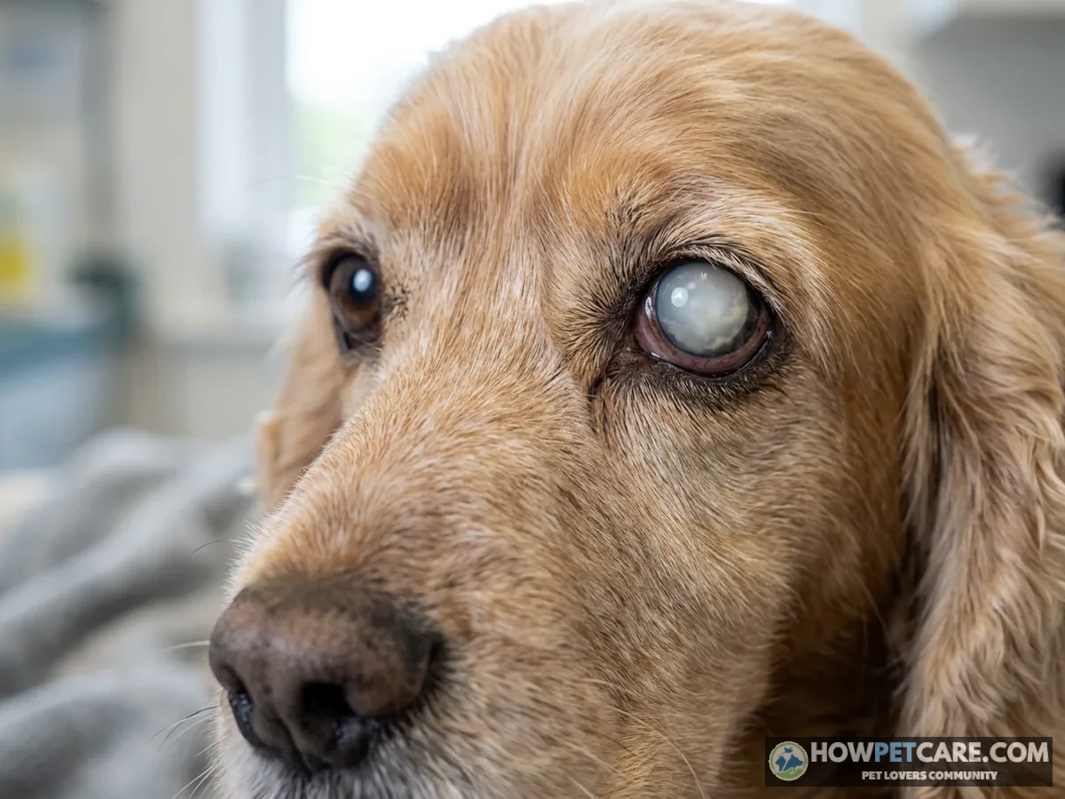

Cataracts in dogs are lens opacities that block light and can impair vision; age-related nuclear sclerosis can look similar but usually preserves sight. Arrange a veterinary exam for new cloudiness or behavior changes. Seek same-day care for pain, redness, swelling, sudden cloudiness, or sudden vision loss.
Cataracts in Dogs: How to Tell if Your Dog’s Cloudy Eyes Need a Vet Now
A cataract is an opacity in the lens that can block light from reaching the retina and, as it grows, impair vision. Nuclear sclerosis, a common age-related lens change, can also make an eye look cloudy but generally preserves vision. You can’t reliably tell the two apart at home.
Arrange a veterinary exam if you notice new cloudiness or changes in your dog’s vision. A red, painful, swollen, or suddenly cloudy eye, or sudden vision loss, needs same-day veterinary attention. These signs can accompany serious conditions, including glaucoma, that require prompt treatment.
If a cataract is confirmed, surgery may restore useful vision in a suitable candidate. Medication can manage inflammation but can’t clear the lens. Surgery is a substantial expense, and the supplied veterinary evidence does not establish a reliable U.S. price range, so ask for an itemized estimate rather than relying on an unverified figure. Recovery commonly starts with intensive medication and activity restrictions, then moves to rechecks and a gradual return to activity. Your ophthalmologist will set the schedule.
The sections ahead cover what to watch for, how cataracts are staged and diagnosed, the questions that turn a vague quote into a realistic surgery estimate, and a week-by-week recovery framework.
Early Signs of Cataracts in Dogs: An At-Home Checklist Before the Vet Visit
A small cataract may not noticeably affect vision. As an opacity grows, a dog may bump into furniture, hesitate on stairs, or have trouble finding familiar objects. Other problems can cause these changes too, so use the checklist to document your concerns, not to diagnose cataracts.
Visual signs you can see
Look at both eyes in ordinary light. Note whether a white, gray, or bluish-white spot appears behind the pupil, whether one eye looks different from the other, and when you first noticed it. A difference such as a drooping lid or a smaller pupil in one eye can point to something like Horner’s syndrome in dogs rather than a lens opacity. A cataract can appear as a white area behind the iris, although not every cataract looks like a solid white disk.

Behavior changes that suggest vision loss
Check any changes you have noticed:
- ☐ Hesitates at stairs, curbs, or when jumping onto familiar furniture.
- ☐ Bumps into door frames or objects, especially after furniture is moved.
- ☐ Seems less confident in dim light or unfamiliar surroundings.
- ☐ Misses a familiar toy or seems to rely more on sound to find it.
- ☐ Startles when approached from the side.
- ☐ Has difficulty locating food or water bowls.
Even one persistent change is worth mentioning to your veterinarian. No validated point score in the supplied evidence can tell you whether your dog has a cataract, or how long it’s safe to wait for an exam.
A 5-minute home vision check
Watch your dog walk through a familiar room in normal light, then note whether behavior changes in dimmer light. Keep the route safe, and don’t set up obstacles that could cause a fall. You can also quietly drop a cotton ball within the dog’s field of view and see whether the eyes follow it. Interest, hearing, scent, and attention can all affect the response, so neither observation proves normal or impaired vision.

Don’t shine a bright light into the eye repeatedly or move your hand toward it to provoke a blink. A veterinarian can assess the pupillary light reflex and other responses safely, and a normal light reflex alone does not rule out a cataract.
Record a short video of any hesitation or bumping, and note when the change began. If you have clear older photos of the eyes, bring those to the appointment too.
Red flags that mean go to a vet today
Seek same-day care for squinting, rubbing, redness, swelling, a bulging eye, new discharge, unequal pupil sizes, or sudden cloudiness or vision loss. A cataract itself isn’t necessarily painful, but inflammation and other eye problems can cause discomfort. If your regular veterinarian can’t assess an urgent eye problem, contact an emergency veterinary service.

A painful or suddenly changed eye needs prompt veterinary assessment, even if your dog still seems able to see.
Cataracts vs. Nuclear Sclerosis: How to Tell the Difference
Side-by-side comparison
Both conditions affect the lens, the structure behind the colored iris. Their appearance can overlap, especially in photographs, so use this comparison to describe what you see, not to make a diagnosis.
| Feature | Cataract | Nuclear sclerosis |
|---|---|---|
| What changes | A lens opacity blocks some light from reaching the retina. | Age-related lens hardening creates a haze while still allowing light through. |
| Typical appearance | May look like a localized white spot or a denser milky-white area behind the pupil. | Often appears as a diffuse bluish-gray haze in an older dog’s eyes. |
| Vision | Depends on the opacity’s size and location; a small cataract may spare vision, while a mature one can cause blindness in that eye. | Generally does not cause blindness and vision is largely preserved. |
| Pattern | May affect one or both eyes and can differ between eyes. | Often appears similar in both eyes. |
| Next step | Veterinary assessment and monitoring; discuss surgery if appropriate. | A veterinary exam to confirm the cause of cloudiness and check for other disease. |

As a rough visual guide, nuclear sclerosis looks like a translucent haze and a dense cataract looks like a more opaque white area. But it’s not a foolproof test. A dog can have both conditions, and a small cataract may be hard to see.
Why a vet exam is needed to confirm
A veterinarian uses light and magnification to examine the lens and determine whether light passes through it normally. A dilated examination or a veterinary ophthalmologist’s slit-lamp exam can help identify the location and extent of an opacity. The vet also checks for associated problems, including inflammation and glaucoma.


What Causes Cataracts in Dogs? Diabetes, Genetics, Age and Other Triggers
Inherited and breed-related cataracts
Inherited disease is a common cause of canine cataracts. Other recognized causes include diabetes mellitus, injury, aging, nutritional imbalance, and chronic inflammation inside the eye.
The veterinary evidence supplied for this guide identifies the following breeds as predisposed, but it does not establish a reliable typical age of onset for each. Inherited cataracts can appear in young dogs, while onset varies with the specific condition and the individual dog. A breed listing does not mean your dog will develop cataracts.
| Breed | Typical onset | Notes |
|---|---|---|
| American cocker spaniel | Variable; breed-specific age not established by the supplied evidence | Listed as predisposed to cataracts. |
| Labrador retriever | Variable; breed-specific age not established by the supplied evidence | Listed as predisposed to cataracts. |
| Poodle | Variable; breed-specific age not established by the supplied evidence | French poodle is listed as predisposed in the veterinary source. |
| Boston terrier | Variable; breed-specific age not established by the supplied evidence | Listed as predisposed to cataracts. |
| Welsh springer spaniel | Variable; breed-specific age not established by the supplied evidence | Listed as predisposed to cataracts. |
Miniature schnauzers, Siberian huskies, bichons frises, border collies, Australian shepherds, and golden retrievers also come up often in discussions of inherited eye disease. The supplied evidence doesn’t establish cataract-specific onset ages for those breeds, so ask a veterinarian or ophthalmologist what screening suits your dog and its family history. Veterinary guidance also advises against breeding affected dogs.

Diabetes and rapid-onset cataracts
Diabetes is a recognized cause of cataracts in dogs. Cloudiness or vision changes in a diabetic dog deserve prompt veterinary assessment, especially if they seem to develop quickly. Tell the vet when you first noticed the change and whether your dog’s diabetes treatment or general health has recently shifted.
Work with your veterinarian on diabetes management and eye monitoring. Good diabetes care matters for overall health, but it doesn’t guarantee a dog will avoid cataracts.
Other causes: injury, inflammation, toxins and age
Eye trauma, chronic uveitis (inflammation inside the eye), and nutritional imbalance can be associated with cataracts, and some cataracts arise with age. An injured or inflamed eye needs veterinary attention whether or not a cataract is visible. Other eye diseases can coexist with a cataract, which is one reason a full examination matters before treatment decisions.
How Dog Cataracts Are Staged and Diagnosed
The four cataract stages
Veterinarians describe cataracts by how much of the lens is affected and whether the lens has changed further. Staging frames the conversation about vision and treatment, but it doesn’t replace a full examination.
| Stage | Lens involvement | Vision impact | Typical management |
|---|---|---|---|
| Incipient | A small portion of the lens is opaque. | Vision is often largely unaffected. | Monitor and discuss how often to recheck. |
| Immature | Part, but not all, of the lens is opaque. | Vision may be reduced, depending on the opacity. | Assess progression and discuss referral if surgery may be an option. |
| Mature | The lens is completely opaque. | The dog can be functionally blind in that eye. | Evaluate eye health, comfort, and surgical candidacy. |
| Hypermature | An advanced cataract has undergone further lens changes. | Vision is severely impaired; inflammation may complicate the condition. | Monitor and treat complications; discuss whether surgery remains appropriate. |

Stage is only one part of the decision. A veterinary source reports that surgery is most successful before a cataract matures, but the right timing depends on the individual eye and the dog’s health. One small cataract may stay stable while another progresses, so your vet can help you plan follow-up.
What the diagnostic exam includes
Your veterinarian will examine the eye with light and magnification and may recommend bloodwork to investigate conditions such as diabetes. Screening may also include checks for uveitis and glaucoma. A veterinary ophthalmologist can perform a more specialized examination, including slit-lamp biomicroscopy.
If surgery is being considered, the specialist may recommend eye-pressure testing, ultrasound to assess structures behind an opaque lens, and an electroretinogram, or ERG, to evaluate retinal function. These tests help determine whether removing the cataract is likely to restore useful vision. Other testing depends on the dog, the eye, and the anesthesia plan.

Treatment for Cataracts in Dogs: Surgery, Eye Drops and Non-Surgical Management
Phacoemulsification is the surgical technique commonly used to remove a cataract. An artificial lens may be implanted. The aim is to restore useful vision, not to promise a perfect result. Whether surgery is worthwhile depends on the health of the eye and retina, the dog’s overall health, the expected benefit, and the family’s ability to manage treatment and cost.
A dog with a healthy retina and manageable medical conditions may be considered for surgery. Uncontrolled inflammation, glaucoma, retinal disease, or other health concerns can change the plan. Diabetes doesn’t automatically rule surgery out, but the veterinary team must consider how safely the dog’s condition can be managed around anesthesia and recovery.
Can eye drops treat cataracts in dogs?
Eye drops prescribed for inflammation can help keep an affected eye comfortable, but they don’t remove an established cataract. Don’t substitute an advertised cataract-clearing drop for a veterinary exam or prescribed treatment. Some eyes need ongoing monitoring even when the dog is adapting well to vision loss.
Natural remedies and supplements: what the evidence supports
| Claim | Practical assessment |
|---|---|
| Lanosterol or similar drops dissolve canine cataracts. | The supplied evidence does not establish that these products reliably clear established cataracts in dogs. |
| N-acetylcarnosine drops reverse cataracts. | The supplied evidence does not establish a reliable reversal benefit in dogs. |
| Antioxidants or supplements prevent or reverse cataracts. | Evidence provided here is insufficient to recommend them for prevention or reversal. Ask your vet before adding supplements. |
| A special diet reverses an existing cataract. | The supplied evidence does not support this claim. A balanced diet remains important for general health. |
| Prescribed anti-inflammatory medication treats associated inflammation. | Medication may be part of a veterinarian’s plan for an inflamed eye; it does not make the cataract disappear. |
| Diabetes management guarantees prevention. | It does not. Follow your vet’s diabetes plan and report eye changes promptly. |
A dog that is coping with poor sight still needs veterinary rechecks. Inflammation or increased eye pressure can threaten comfort even when surgery is not planned.
Living with a blind or partially blind dog
Many dogs adapt to gradual vision loss, particularly in familiar surroundings. Keep furniture, food, and water in consistent places. Block access to stairs or pools, and use verbal cues before approaching or guiding your dog. A harness and a predictable walking route can help outside. Don’t introduce obstacles just to test vision.

Non-surgical management is an active care plan, not a cure. Ask your veterinarian which signs should prompt a recheck, whether eye-pressure monitoring is needed, and whether medication is appropriate for inflammation or discomfort.
Dog Cataract Surgery Cost: Realistic Price Ranges and What Moves the Price
Typical cost range and what is included
Cataract surgery can cost several thousand dollars, but the supplied evidence does not verify a specific U.S. price range or individual line-item prices. A numerical estimate without a supporting source could give you a misleading budget. Request a written estimate for your dog’s particular workup, surgery, and aftercare, and confirm whether the amounts cover one eye or both.
A useful estimate separates the consultation, diagnostic testing, anesthesia, procedure, artificial lens if used, medications, and rechecks. Some practices bundle services that others bill separately. A lower initial figure isn’t necessarily cheaper once the full care plan is included.

Factors that raise or lower the price
What you pay can depend on location, the specialty practice, whether one or both eyes are treated, the tests required, and the dog’s health. Extra monitoring, complications, additional medication, or further procedures can increase the total.
When you ask for estimates, compare these scenarios instead of assuming they cost the same:
- One uncomplicated eye: Ask what the quoted procedure includes and which tests, medications, and rechecks are separate.
- Both eyes: Ask whether the quote covers both procedures, both implants if appropriate, and the complete follow-up plan.
- A more complex case: Ask how diabetes, inflammation, abnormal pressure, or another eye condition could change the workup and estimate.
Before committing, ask:
- Are the ERG, ultrasound, bloodwork, anesthesia, and lens implant included?
- How many rechecks and how much medication are included, and what is billed separately?
- If testing shows that surgery is not advisable, what charges will already apply?
- What is the plan if glaucoma, retinal detachment, or another complication requires additional care?
If you have pet insurance, check the policy’s terms for hereditary conditions, waiting periods, and pre-existing conditions before assuming coverage. Ask the veterinary practice about payment arrangements or financing if needed.
Is cataract surgery worth it?
Surgery may be worth considering when testing suggests vision can be restored, the dog can safely undergo anesthesia, and the family can manage the expense and aftercare. It may be less suitable when another eye disease limits the likely benefit, medical risks are substantial, or the required treatment schedule isn’t manageable. A comfortable dog can still enjoy a good quality of life with vision loss.
Recovery After Dog Cataract Surgery: Week-by-Week Timeline and Medication Schedule
Recovery varies with the dog, the operation, and any complications. Treat the following as a planning framework, not a prescription. Your ophthalmologist’s instructions always take precedence, including the names, doses, timing, and order of medications.
| Period | Medication and rechecks | Activity and care |
|---|---|---|
| First 48 hours | Expect a potentially intensive drop schedule and a prompt postoperative check as directed. Some dogs also receive oral medication. | Keep the protective cone on as instructed; use a leash for short bathroom trips and prevent rubbing. |
| Weeks 1–2 | Continue drops exactly as prescribed. The vet may adjust the schedule at a recheck if healing permits. | Restrict activity; avoid rough play and use a harness if advised. Do not remove the cone early without approval. |
| Weeks 3–6 | Attend scheduled rechecks; medication may be reduced or continued according to the exam. | Increase activity only when the ophthalmologist clears it. |
| Longer term | Some dogs need extended medication and periodic eye examinations. | Report new redness, discomfort, cloudiness, or vision changes promptly. |

The first 48 hours
Plan for someone to give medication and watch the dog closely. Discharge instructions may include antibiotic and anti-inflammatory eye drops, and sometimes medication for eye pressure or pain. The exact combination and frequency are individual. Phone reminders and a written log help household members avoid missed or duplicated doses.
Weeks 1–2: the critical window
Keep the e-collar in place according to the surgeon’s instructions, including during sleep if directed. Limit walks to those the care team approves, prevent rough play, and attend the scheduled recheck even if the eye looks good. Keep outings short in hot weather too, since dogs cool down mainly by panting rather than sweating and can overheat quickly when restricted. Never taper or stop drops on your own. Call the clinic if a dose is missed or your dog won’t tolerate treatment.
Weeks 3–6 and long-term care
The ophthalmologist may gradually reduce medication and allow more activity as healing progresses. Some dogs need anti-inflammatory drops for an extended period, and long-term rechecks help detect problems such as glaucoma. Ask for a written follow-up plan that includes expected costs and who to contact outside office hours.
Complications to watch for
Contact the surgical team promptly about increased redness, squinting, rubbing, swelling, discharge, a newly cloudy eye, or worsening vision. Glaucoma and retinal detachment are among the complications the veterinary team monitors for. If your dog seems painful or suddenly loses vision and you can’t reach the surgeon, seek urgent veterinary care. Apply the same urgency to sudden non-eye emergencies, such as paraphimosis or urinary obstruction in male dogs.
Your Next Steps: A Decision Guide for Dogs With Suspected Cataracts
- Document what changed. Use the checklist, note the timeline, and bring a short video or earlier eye photos if available.
- Arrange an exam. Seek same-day care for pain, sudden cloudiness, or sudden vision loss. Tell the clinic promptly if your dog has diabetes.
- Ask what the diagnosis means. If a cataract is confirmed, ask about its stage, whether the other eye is affected, and whether referral to a veterinary ophthalmologist is appropriate.
- Request a written plan and estimate. Clarify testing, likely benefit, anesthesia risks, medication, rechecks, and what happens if complications arise.
- Plan ongoing care. Whether or not you pursue surgery, agree on rechecks and changes that will help your dog get around safely.
Decision checklist: surgery or non-surgical management
Discuss surgery if the specialist expects useful vision to return, the dog is a reasonable anesthesia candidate, and you can carry out the medication and recheck schedule. Discuss non-surgical management if testing suggests limited benefit, the risks or cost are too high, or aftercare isn’t feasible. In either case, ask how the team will monitor comfort and eye pressure, what changes require urgent care, and when to revisit the decision.
| Stage | Lens Involvement | Vision Impact | Typical Management | Surgical Candidate Status (Inferred) |
|---|---|---|---|---|
| Incipient | A small portion of the lens is opaque. | Vision is often largely unaffected. | Monitor and discuss how often to recheck. | Unlikely surgical candidate at this stage unless rapid progression is expected, as vision is largely unaffected (surgery usually discussed or performed as it progresses, most successful before maturing). |
| Immature | Part, but not all, of the lens is opaque. | Vision may be reduced, depending on the opacity. | Assess progression and discuss referral if surgery may be an option. | Ideal / strong surgical candidate; surgery is often reported to be most successful before the cataract fully matures. |
| Mature | The lens is completely opaque. | The dog can be functionally blind in that eye. | Evaluate eye health, comfort, and surgical candidacy. | Potential surgical candidate if the retina and overall eye health remain suitable (requires evaluation of retinal function to determine if vision can be restored), though success rates may be higher if operated on earlier. |
| Hypermature | An advanced cataract has undergone further lens changes. | Vision is severely impaired; inflammation may complicate the condition. | Monitor and treat complications; discuss whether surgery remains appropriate. | Guarded / complex surgical candidate; surgery may be less suitable or complicated due to advanced changes, intraocular inflammation, secondary eye conditions, and potential risks. |
FAQ
Can diabetic dogs get cataracts?
Yes. Diabetes is one of the most common causes of cataracts in dogs, and the lens opacity can develop quickly, sometimes within weeks to months of diagnosis. Keeping blood sugar well controlled and having regular eye exams helps you catch changes early.
Do cataracts in dogs go away on their own?
No, true cataracts do not resolve by themselves and may progress over time. Some small or incipient cataracts stay stable for years, which is why regular monitoring matters.
Is a dog with cataracts in pain?
The cataract itself is usually not painful, but complications such as lens-induced uveitis or glaucoma can cause pain, redness and squinting. Any sign of discomfort in the eye needs prompt veterinary attention.
Can a blind dog with cataracts still have a good quality of life?
Many dogs adapt well to vision loss, especially in a familiar home with a consistent layout and verbal cues. Their other senses compensate, and regular vet checks can ensure the eyes stay comfortable.
At what age do dogs get cataracts?
It depends on the cause. Hereditary cataracts can appear in young dogs, sometimes before age 3, while age-related cataracts are more common in middle-aged and senior dogs. Diabetic cataracts can appear at any age after the disease develops.