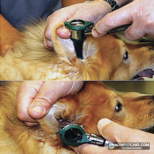

A yeast infection in dogs, caused by an overgrowth of Malassezia yeast, shows up as greasy, dark crusts, a strong “corn‑chip” odor, and intense itching—often on ears, paws, or skin folds. Prompt veterinary testing confirms the diagnosis and guides topical or oral antifungal treatment.
A yeast infection in dogs, clinically termed Malassezia dermatitis, happens when the skin yeast Malassezia pachydermatis multiplies out of control. The result is greasy, inflamed, and distinctly foul‑smelling skin or ears. This organism naturally inhabits canine skin; it is not contagious. Instead, overgrowth flags an underlying breach in the dog’s epidermal barrier or immune defenses. If your dog has a persistent "corn‑chip" odor, intense pruritus, or oily, dark crusts around the paws, ears, or skin folds, schedule a veterinary visit. Prompt clinical testing identifies the root cause and keeps the condition from becoming an entrenched, chronic issue.
Signs of a Yeast Infection in Dogs: Identifying Malassezia by Body Site
Catching yeast dermatitis early makes treatment far more straightforward. The organism thrives in warm, sheltered microenvironments—primarily the ear canals, interdigital spaces of the paws, and deep body folds.
- Musty “corn‑chip” odor that persists shortly after a thorough bath.
- Greasy, brown‑black crusts or waxy discharge that feel slick to the touch.
- Hyperpigmentation and thickened, “elephant‑skin” appearance across inflamed patches.
- Intense itching or licking, often localized to the site.
Dog Ear Yeast Infection Symptoms vs. Paw and Fold Dermatitis
| Body Site | Typical Appearance | Common Odor | Typical Behavior |
|---|---|---|---|
| Ears (otitis externa) | Dark brown or black debris within the canal; reddened ear margin; possible swelling | Strong, sour, “musty” smell | Head shaking, scratching at the base of the ear |
| Paws (interdigital) | Redness between toes, scaling, sometimes a shiny sheen; may see small crusts | Musty or slightly sweet odor | Licking or chewing of the pads, reluctance to walk |
| Skin folds (face, tail, underbelly) | Macerated, wet‑looking skin; thickened, hyperpigmented folds; sometimes ulceration | Persistent musty odor, especially after activity | Scratching, rubbing against furniture, “fold‑licking” |
Visual cues and characteristic odors point toward Malassezia, but they cannot provide a definitive diagnosis on their own. Yeast overgrowth frequently pairs with atopic allergies or secondary bacterial infections, making in‑clinic microscopic evaluation essential.
Visual Changes: Hyperpigmentation, Greasy Seborrhea, and “Elephant Skin”
As Malassezia populations spike, their metabolic by‑products irritate the epidermis. This chronic stimulation accelerates keratinization and spikes sebum production. Over time, the skin develops lichenification—the thickened, darkened, cobblestone texture colloquially called "elephant skin." The oily coat feel comes directly from the yeast feeding on cutaneous lipids, not from dietary carbohydrates.

Yeast Infection vs. Bacterial Infection, Allergies, and Mange
Because redness and relentless itching accompany many canine dermatoses, misidentifying yeast dermatitis is easy. The matrix below highlights key diagnostic differences.
| Condition | Primary Cause | Distinctive Lesions & Exudate | Odor Profile | Definitive Diagnostic Test |
|---|---|---|---|---|
| Yeast Dermatitis (Malassezia) | Opportunistic overgrowth of normal skin yeast | Greasy, brown‑black crusts; hyperpigmented, thickened “elephant skin”; macerated folds | Musty, “corn‑chip” smell | Acetate tape or skin‑scrape cytology > 5 organisms/HPF |
| Bacterial Pyoderma | Overgrowth of Staphylococcus spp. (often secondary) | Pustules, honey‑colored crusts, erythema; may be oozing | Often foul but not the characteristic “musty” | Cytology showing neutrophils with intracellular cocci |
| Atopic Allergies / Food‑Induced Dermatitis | Environmental or dietary hypersensitivity | Lichenification, erythema, pododermatitis; no specific crust | Usually no odor or mild stale odor | Cytology may show ≤5 yeast/HPF; diagnosis via intradermal/allergy testing |
| Sarcoptic / Demodectic Mange | Mite infestation (Sarcoptes or Demodex) | Papules, crusts, alopecia, intense pruritus; secondary bacterial infection possible | Typically no distinct odor | Skin scrape revealing live mites (or mite count) |
Why laboratory testing matters – The lesions of yeast dermatitis overlap with bacterial pyoderma and allergic dermatitis, and coinfections are common. A simple skin‑scrape or acetate tape impression examined under a microscope tells the clinician how many yeast cells are present, guiding appropriate therapy.

What Causes Yeast Infections in Dogs? The Secondary Overgrowth Mechanism
Malassezia pachydermatis harmlessly colonizes canine skin and external ear canals under normal physiological conditions[ #1]. Clinical disease begins when local microclimates or systemic shifts allow the yeast population to surge.
- Moisture retention in skin folds, between paw pads, or after swimming creates a warm, anaerobic niche.
- Barrier disruption from allergies, hormonal imbalances (hypothyroidism, Cushing’s disease), or repetitive trauma allows yeast to invade deeper layers.
- Immune dysregulation – Dogs with atopic dermatitis or food‑induced hypersensitivity often have an overreactive immune response that paradoxically reduces the skin’s ability to control microbial growth.
Anatomical Risk Factors: Deep Skin Folds and Pendulous Ear Canals
Conformation plays a direct role. Breeds with prominent skin rolls (such as Basset Hounds and English Bulldogs) or pendulous, heavy pinnae (such as Cocker Spaniels and West Highland White Terriers) naturally trap moisture and shed cells. These microenvironments encourage rapid Malassezia proliferation, and several of these breeds also inherit structural defects in their epidermal lipid barrier.

Systemic Drivers: Allergies, Immune Dysfunction, and Endocrine Disease
Underlying allergies represent the most frequent primary trigger for recurrent yeast dermatitis[ #2]. Both environmental allergens (pollen, house dust mites) and cutaneous adverse food reactions compromise skin defenses. Endocrine diseases such as hypothyroidism and hyperadrenocorticism (Cushing’s disease) also undermine local immunity and epidermal turnover. Severe cases frequently see flares recurring 2–3 times per year until the systemic disease is controlled.
Veterinary Triage: When to Schedule a Routine Visit vs. Seek Immediate Care
While mild itching rarely warrants emergency intervention, certain clinical signs demand immediate veterinary evaluation.
Veterinary Triage Checklist for Yeast‑Related Dermatitisnn- ☐ Mild, localized itching (paws or skin folds) with no open wounds → schedule a routine appointment within 3–5 days.
- ☐ Persistent “corn‑chip” odor but lesions are dry and not painful → routine visit.
- ☐ Ear discharge that is dark brown, malodorous, and the dog shakes its head but no pain → routine appointment.
- ☐ Sudden severe ear pain, head tilt, nystagmus, or loss of balance → seek emergency veterinary care now.
- ☐ Deep ulcerations, bleeding, or rapid swelling of a skin fold → urgent evaluation (same‑day).
- ☐ Signs of systemic illness (lethargy, fever, vomiting, weight loss) alongside skin issues → immediate vet visit.
- ☐ Uncontrolled diabetes or Cushing’s disease with new yeast flare‑up → prioritize urgent endocrine work‑up.
nIf any red‑flag box is checked, call your veterinarian or an emergency clinic immediately.
Red‑Flag Signs that Require Immediate Attention
- Intense ear pain – your dog vocalizes, snaps, or pulls away sharply when the pinna or canal is touched.
- Neurologic signs – head tilts, ataxia, or abnormal eye movements (nystagmus) point toward middle or inner ear involvement.
- Rapidly expanding swelling or ulceration – suggests aggressive secondary bacterial cellulitis or deep tissue compromise.
If your dog shows any of these signs, avoid home rinses and contact an emergency veterinary hospital immediately.

How Veterinarians Diagnose Canine Yeast Infections and Typical Costs
Diagnostic Techniques
- Acetate tape impression – a clear adhesive strip pressed onto the skin or ear canal, then examined under a microscope. It quantifies yeast organisms per high‑power field.
- Skin‑scrape cytology – a sterile blade gently scrapes the lesion; the sample is stained and viewed for yeast, bacteria, and inflammatory cells.
- Ear swab cytology – similar to tape impression but performed within the ear canal to differentiate yeast from bacterial otitis.
- Culture (rarely needed) – ordered when the infection does not respond to standard therapy, to identify resistant organisms.
- Blood work – thyroid panels or adrenal testing may be recommended if underlying endocrine disease is suspected.
These in‑clinic tests take only a few minutes, yet they yield the objective microscopic confirmation needed to prescribe targeted antifungals rather than guessing.
Cost Breakdown (2026 US dollars)
| Service or Medication | Purpose | Typical Cost Range (USD) | Frequency |
|---|---|---|---|
| Initial veterinary exam | General assessment, sample collection | $50–$100 | One‑time |
| Cytology (each site) | Microscopic identification of yeast | $30–$60 | Usually 1–3 sites per visit |
| Medicated shampoo (e.g., chlorhexidine‑ketoconazole) | Topical antifungal therapy | $30–$45 (8‑oz bottle) | 2‑4 weeks, then as maintenance |
| Oral antifungal (e.g., ketoconazole) | Systemic control for generalized infection | $80–$120 (30‑day supply) | 4–12 weeks, with liver monitoring |
| Follow‑up re‑check | Evaluate response, adjust therapy | $30–$50 | 2–3 weeks after start |
| Allergy testing or elimination diet trial | Identify underlying trigger | $150–$300 | 8–12 weeks |
These estimates reflect broad clinical averages. Total expenses shift depending on your geographic location, your dog’s size, and the diagnostic path chosen.
Veterinary Medical Treatment: Topical Therapies vs. Oral Antifungals
Understanding the clinical difference between prescription antifungal therapy and supportive home care is essential for successful management: prescription antifungal therapy directly inhibits fungal replication and destroys yeast cell membranes, whereas supportive home care focuses on mechanical cleansing, removing sebum and debris, and drying moisture to minimize environmental recurrence.
First‑Line Topical Options
- Chlorhexidine‑ketoconazole shampoo – combines a broad‑spectrum antiseptic with an azole antifungal. The shampoo is applied to the affected area and left in place for 5–10 minutes to allow penetration of the yeast cell wall (minimum 10 minutes is supported by in‑vitro studies).
- Miconazole wipes or sprays – useful for quick spot treatment of paws and folds; they dry rapidly and reduce the need for frequent bathing.
- Veterinary ear suspensions (e.g., a combination of miconazole, polymyxin B, and hydrocortisone) – administered directly into the ear canal to target otitis externa.

When Oral Antifungals Are Needed
Systemic drugs such as ketoconazole, itraconazole, fluconazole, or terbinafine are reserved for:
- Generalized skin involvement covering > 30 % of the body surface.
- Refractory cases that do not improve after 2–3 weeks of topical therapy.
- Deep infections involving the ear canal or nail beds.
Oral azoles undergo hepatic metabolism and carry potential hepatotoxicity risks. Clinicians monitor serum liver enzymes (ALT, AST) every 2–4 weeks throughout systemic treatment[ #3]. Therapy often continues for several consecutive months to clear deep‑seated reservoirs in the hair follicles and nail beds.
Expected Timeline for Resolution
- Topical therapy alone – noticeable reduction in itching within 7 days, with visible clearing of lesions in 2–4 weeks.
- Oral therapy – itching may improve within the first week, but full skin normalization can require 6–12 weeks, plus an additional 1–2 weeks of maintenance to prevent relapse.
Safe Supportive Home Care: Bathing Contact Times, Paw Drying, and Ear Administration
-
Gather Supplies
Gather a medicated chlorhexidine‑ketoconazole shampoo, a bucket of warm (≈38 °C/100 °F) water, soft towels, cotton balls, and the prescribed ear suspension.
-
Pre‑Bath Preparation
Brush the coat to remove loose debris. Pat the paws and skin folds dry with a towel to reduce excess moisture before bathing.
-
Apply Shampoo
Wet the affected area thoroughly, then lather the medicated shampoo. Set a timer for 10 minutes and keep the lather on the skin for the full dwell time (minimum 10 minutes) to achieve optimal antifungal penetration
-
Rinse Completely
Use lukewarm water to rinse all shampoo residue; remaining product can irritate compromised skin.
-
Immediate Drying
Gently pat the area dry. For deep folds, press a second clean towel into the crease for 30 seconds to extract hidden moisture.
-
Ear Medication
Tilt the dog’s head slightly upward, fill the ear canal with the prescribed drops (usually 3–5 drops). Massage the base of the ear for 20 seconds to spread the medication, then allow the dog to shake its head for a few seconds. Wipe excess solution from the outer ear with a clean cotton ball—do not insert cotton swabs deep into the canal.
-
Post‑Treatment Observation
Watch for signs of pain or swelling. If the ear canal feels unusually painful, stop cleaning and contact your veterinarian immediately.
-
Frequency & Record Keeping
Perform this protocol twice weekly for the first two weeks, then once weekly until lesions are fully resolved. Keep a simple log of dates, shampoo dwell time, and any changes in odor or itch level to share with your vet.
Tip: After each bath, use a dry, lint‑free cloth to fold‑dry skin folds. Press the cloth into the fold for a few seconds; this removes moisture that would otherwise become a breeding ground for yeast.

The Truth About Home Remedies and ‘Anti‑Yeast’ Diets
Myth 1 – Apple Cider Vinegar (ACV) cures yeast infection
- Fact: ACV is highly acidic. Applying it to inflamed, macerated skin can strip the protective lipid barrier, cause stinging, and alter skin pH in a way that actually encourages Malassezia growth. Use only under veterinary direction.
Myth 2 – Coconut oil “starves” yeast because of lauric acid
- Fact: While lauric acid has in‑vitro antifungal activity, on compromised skin it adds a fatty substrate that Malassezia loves to feed on. Topical coconut oil can worsen greasy lesions and delay healing.
Myth 3 – Cutting dietary carbs “starves” skin yeast
- Fact: Malassezia obtains nutrients from skin lipids, not from dietary carbohydrates. Low‑carb or “yeast‑starving” diets have no proven effect on skin overgrowth. Effective dietary management focuses on identifying true food allergens via a supervised elimination diet, not on carbohydrate restriction.
Why elimination diets work (and low‑carb diets don’t)
A structured veterinary elimination diet uses a novel-protein or hydrolyzed-protein diet exclusively for 8–12 weeks, cutting out suspect antigens. If the patient’s skin calms and yeast stops recurring, an adverse food reaction was the underlying driver. This diagnostic method relies on controlled clinical immunology, unlike retail "anti-yeast" kibbles that lack empirical backing.
Action Plan: Preventing Recurrent Malassezia Overgrowth in Dogs
Daily Moisture Management
- After swimming or a rainy walk, thoroughly dry the paws, especially between the toes, using a clean towel.
- Fold‑dry any skin creases (e.g., under the belly, neck, tail base) by gently pressing a dry cloth into the fold for at least 30 seconds.
- Inspect ears daily for moisture or discharge; gently wipe the outer ear with a cotton ball if needed.
Proactive Maintenance Regimen
- Weekly antiseptic wipes (chlorhexidine‑based) on high‑risk areas can keep yeast counts low without over‑drying the skin.
- Maintenance baths with a low‑dose medicated shampoo once a month help prevent flare‑ups in predisposed breeds.
Long‑Term Management of Underlying Triggers
- Work with your veterinarian to identify and control allergies – options include sub‑cutaneous allergen immunotherapy, cytokine‑targeted drugs (e.g., Apoquel), or regular bathing with antiseptic shampoo.
- Monitor endocrine health – regular thyroid panels and adrenal assessments can catch hypothyroidism or Cushing’s early, reducing the risk of recurrent yeast infections.
- Weight management – overweight dogs have deeper skin folds and reduced skin ventilation, both of which favor yeast growth.
Pairing consistent home management with targeted medical protocols gives dogs long-term relief from Malassezia overgrowth. If you spot greasy crusting or that telltale corn‑chip scent, reach out to your veterinarian for definitive diagnostic testing and an appropriate prescription plan.


| Condition | Primary Cause | Distinctive Lesions & Exudate | Odor Profile | Definitive Diagnostic Test |
|---|---|---|---|---|
| Yeast Dermatitis (Malassezia) | Opportunistic overgrowth of normal skin yeast (Malassezia) | Greasy, brown‑black crusts; hyperpigmented, thickened “elephant skin”; macerated folds | Musty, “corn‑chip” odor | Cytology (acetate tape or skin scrape) showing >5 organisms/HPF |
| Bacterial Pyoderma | Overgrowth of Staphylococcus spp. (often secondary) | Pustules, honey‑colored crusts, erythema; may have oozing exudate | Foul odor (lacks characteristic musty note) | Cytology showing neutrophils with intracellular cocci |
| Atopic Allergies / Food‑Induced Dermatitis | Environmental or dietary hypersensitivity | Erythema, lichenification, pododermatitis; no specific crust or exudate | Mild stale odor or no distinct odor | Intradermal allergy testing or elimination diet trials (cytology may show ≤5 yeast/HPF) |
| Sarcoptic / Demodectic Mange | Mite infestation (Sarcoptes or Demodex) | Papules, crusts, alopecia, intense pruritus; secondary bacterial infection possible | No distinct odor | Deep or superficial skin scrape revealing live mites |
FAQ
Can I use human Monistat or Lotrimin on my dog’s yeast infection?
While human topical antifungals like miconazole or clotrimazole contain similar active ingredients, using them without veterinary guidance is risky. Dogs frequently lick off topical creams, causing gastrointestinal distress, and human formulations lack the specific vehicle and antimicrobial combinations needed to treat canine multi-organism infections.
Is a dog yeast infection contagious to humans or other pets?
No, Malassezia yeast infections are not contagious. The yeast is a normal resident organism already present on healthy canine skin and only overgrows when an individual animal’s skin microclimate, immune system, or skin barrier becomes compromised.
Can giving my dog probiotics clear up skin yeast infections?
Probiotics support overall gastrointestinal and immune health, but there is no clinical evidence that oral probiotics alone will eliminate an active cutaneous Malassezia overgrowth. Active yeast dermatitis requires direct topical or systemic antifungal therapy prescribed by a veterinarian.
How long does it take for a dog’s yeast infection to fully heal?
Mild localized yeast infections typically improve within 7 to 14 days of consistent treatment, while generalized or chronic skin fold infections often require 3 to 4 weeks. Treatment must always continue for 7 to 10 days past the complete resolution of visible symptoms to prevent immediate relapse.