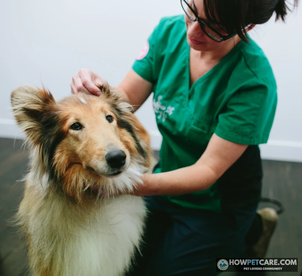

Keratoseborrhea in dogs is a chronic dermatological disorder of abnormal skin cell turnover (cornification) and altered sebum production. Instead of a normal 21-day cycle, epidermal cells mature in 3 to 7 days, causing either dry scaling (seborrhea sicca) or excessive grease and odor (seborrhea oleosa), usually secondary to allergies, parasites, or endocrine disease.
In canine dermatology, keratoseborrhea is a functional defect of cornification and sebum production where a dog’s skin sheds immature epidermal cells too rapidly while producing abnormal levels of cutaneous lipids. Pet owners researching this condition often hit an immediate wall of confusion: online search engines frequently conflate canine keratoseborrhea with human seborrheic keratosis. These two medical conditions are entirely unrelated. Human seborrheic keratosis describes benign, warty, non-cancerous epidermal growths that commonly emerge after age 50 and measure over 1 inch (2.5 cm) across. By contrast, canine keratoseborrhea is not a skin tumor; it is an extensive derangement of skin cell maturation that leaves your pet with foul-smelling, intensely greasy, or severely flaky skin.
Over 80% of dogs displaying seborrheic signs do not suffer from an incurable genetic flaw. Instead, they exhibit secondary seborrhea—an outward cutaneous reaction provoked by underlying allergic dermatitis, endocrine disorders, or microscopic skin parasites. Resolving the condition permanently requires identifying that underlying trigger through a structured veterinary workup while aggressively restoring the epidermal barrier using targeted topical pharmacology.
What Is Canine Keratoseborrhea? (And What the SERP Gets Wrong)
Keratoseborrhea (often called canine seborrhea or a keratoseborrheic disorder) is a clinical syndrome characterized by defective keratinization and altered sebaceous gland activity. Understanding this condition begins with the structure of canine skin.
The Epidermal Turnover Cycle Explained
The outermost layer of a dog’s skin, the stratum corneum, operates on a tightly calibrated cellular schedule. In healthy dogs, basal cells divide in the lowest layer of the epidermis, slowly mature as they produce keratin, migrate upward, die, and slough off invisibly. This normal epidermal turnover cycle takes approximately 21 days.
When a dog develops a keratoseborrheic disorder, this process derails completely. The epidermal transit time accelerates dramatically, dropping from 21 days down to just 3 to 7 days. Because cellular turnover speeds up so aggressively, basal cells divide faster than they can properly cornify. Instead of shedding invisible individual dead cells, the skin casts off massive sheets of immature, sticky keratinocytes glued together by altered lipids. Simultaneously, the sebaceous glands alter their sebum production—either shutting down to leave brittle scales or hyper-secreting abnormal free fatty acids that saturate the coat canopy.

Canine Keratoseborrhea vs. Human Seborrheic Keratosis
Because medical terminology sounds similar, automated search algorithms regularly confuse pet owners by serving articles from human clinical databases. Clarifying the difference prevents unnecessary panic:
- Human Seborrheic Keratosis: A benign, non-cancerous cutaneous neoplasm. In humans, these present as discrete, raised, waxy or scaly plaques that look "pasted onto the skin" on the chest, face, or back. They do not represent a widespread disease of skin cell turnover or sebum chemistry.
- Canine Keratoseborrhea: A generalized disorder of cornification (keratinization) and oil production. It affects large body regions—often spanning the entire dorsal spine, paws, armpits, and ears—causing diffuse scaling, follicular casts, and profound barrier compromise.
When this protective epidermal barrier breaks down, the skin loses moisture via elevated trans-epidermal water loss (TEWL) and becomes vulnerable to opportunistic colonization by bacteria and yeast.
Classifying the Condition: Seborrhea Sicca vs. Seborrhea Oleosa
Veterinarians classify canine seborrhea along a physical spectrum ranging from dry and powdery to profoundly greasy and exudative. Correctly identifying where your dog sits on this spectrum determines which medicated shampoo actives will resolve the issue and which will make it worse.
Seborrhea Sicca: Characteristics of Dry Scaling
Seborrhea sicca is the dry manifestation of cornification dysfunction. In this form, epidermal hyperproliferation occurs alongside inadequate or biochemically altered sebum, stripping the skin of essential hydration.
- Clinical presentation: The coat appears dull, lifeless, and bleached of luster. Hair shafts are brittle and break easily.
- Scale morphology: Owners notice fine, chalky, non-adherent white-to-slate-gray flakes distributed freely throughout the fur and littering the dog’s bedding—resembling heavy human dandruff.
- Distribution: Primarily affects the dorsal trunk, running from the neck down the spine to the base of the tail, as well as the outer margins of the ear pinnae.
- Pruritus (itching): Generally mild to moderate, driven primarily by xerosis (severe skin dryness) rather than rampant microbial overgrowth.
Seborrhea Oleosa: Greasy Crusts and Rancid Odor
Seborrhea oleosa represents the opposite extreme: rampant overproduction of sebaceous lipids combined with accelerated keratinocyte shedding.

- Clinical presentation: The skin feels slick, clammy, and intensely greasy to the touch. Petting the dog leaves an oily, yellowish lipid film on human fingers.
- Scale morphology: Rather than dry flakes, the skin generates thick, adherent, yellow-brown crusts that cake against the skin surface and glue adjacent hair shafts into greasy clumps.
- Follicular plugging: Hyperkeratosis inside the hair follicle produces prominent comedones ("blackheads") and follicular casts—thick cuffs of keratin and sebum that wrap tightly around the base of hair shafts.
- Odor profile: Severe, pervasive, and rancid. When sebaceous glands overproduce lipids, local cutaneous bacteria and yeast hydrolyze those triglycerides into volatile free fatty acids, creating an unmistakable "spoiled butter" or stale corn-chip smell.
- Pruritus: Severe and unremitting, often provoking intense self-trauma, excoriations, and chronic licking.
Mixed Seborrhea: When Dry and Oily Presentations Overlap
Pure presentations represent clinical anchors; many canine patients actually present with mixed keratoseborrheic disorders. A classic mixed presentation features dry, chalky seborrhea sicca scaling along the dorsal spine, alongside severe, greasy seborrhea oleosa within intertriginous friction zones (the armpits, groin, ventral neck folds, and between the toes).
| Clinical Feature | Seborrhea Sicca (Dry) | Seborrhea Oleosa (Oily) |
|---|---|---|
| Skin & Coat Feel | Dull, lacklustre coat; dry, brittle hair shafts; powdery to the touch. | Greasy, slick skin; clumpy, wet-feeling coat that leaves residue on hands. |
| Scale Morphology | Fine, chalky, white-to-slate-gray scales that flake freely off the hair. | Thick, adherent yellow-to-brown lipid crusts that glue hair clumps together. |
| Follicular Plugs | Rare; primarily superficial stratum corneum micro-scaling. | Marked comedones ("blackheads") and follicular casts (collars of scale around hairs). |
| Odor Profile | Minimal to non-existent; faint dusty smell. | Pungent, rancid, "spoiled butter" odor driven by oxidized free fatty acids. |
| Primary Anatomical Sites | Dorsal backline, flanks, neck, pinnae margins. | Ventral abdomen, axillae (armpits), inguinal folds, peri-anal zone, nail beds. |
| Microbial Complications | Mild transient superficial pyoderma; low yeast load. | Severe secondary Malassezia (yeast) dermatitis and Staphylococcus pyoderma. |
| Front-Line Active Ingredients | Micronized sulfur (2%), salicylic acid (2%), ceramides. | Benzoyl peroxide (2.5–3%), selenium sulfide (1%), chlorhexidine (3–4%). |
Never apply human anti-dandruff shampoos (such as Head & Shoulders or Selsun Blue formulated for human scalps) to a dog. Human skin has an acidic pH of 4.5 to 5.5, whereas canine skin is significantly more neutral to alkaline, averaging 6.2 to 7.5. Washing a dog with human formulations strips their delicate stratum corneum, worsens epidermal turnover defects, and induces acute chemical dermatitis.
Underlying Causes: Primary Hereditary Defects vs. Secondary Triggers
To manage canine seborrhea successfully, you must determine whether the condition is an inherited, primary defect in the skin’s blueprint or an acquired, secondary symptom triggered by an underlying systemic pathology.
Primary Idiopathic Seborrhea and Breed Predispositions
Primary keratoseborrhea is an inherited, congenital disease where the abnormal 3-to-7-day epidermal turnover rate occurs intrinsically without any external disease driving it. Because it is encoded in the dog’s DNA, primary seborrhea manifests early in life—typically between 6 and 18 months of age—and progresses in severity throughout the dog’s life.
| Breed | Typical Age of Onset | Predominant Phenotype | Hallmark Clinical Lesions |
|---|---|---|---|
| American Cocker Spaniel | 3 to 12 months | Severe Seborrhea Oleosa | Pronounced rancid odor, proliferative ceruminous otitis externa, interdigital greasy casts, plaque-like ventral crusts. |
| West Highland White Terrier | 6 to 18 months | Mixed / Lichenified | Progressive dark hyperpigmentation, severe chronic pruritus, secondary Malassezia colonization of axillae and groin. |
| Basset Hound | 6 to 12 months | Pronounced Oleosa with Intertrigo | Heavy exudative scaling within deep skin folds, prominent follicular casts, greasy ceruminous debris along pendulous ears. |
| English Springer Spaniel | 1 to 2 years | Seborrhea Sicca to Mixed | Extensive chalky scaling over the dorsal lumbar region, moderate follicular plugging, dry brittle hair shafts. |
| Golden Retriever | 2 to 4 months (ichthyosis variant) | Severe Non-Inflammatory Sicca | Very large, dark parchment-like sheet scales over flanks and ventrum without odor or severe scratching. |

Endocrine and Metabolic Triggers
In older dogs (generally those over 4 to 5 years old) that suddenly develop scaling and greasy coats without a youthful history of skin issues, secondary seborrhea driven by an endocrine disorder is the primary suspect. Hormones regulate basal keratinocyte proliferation and sebaceous gland output:
- Hypothyroidism: Thyroid hormone acts as the metabolic throttle for cellular division in the skin. When thyroid hormone drops, epidermal turnover slows pathologically, hair follicles arrest in the resting (telogen) phase, and the stratum corneum fails to desquamate properly, producing thick, dry flakes, symmetrical flank alopecia, and secondary oily seborrhea.
- Hyperadrenocorticism (Cushing’s Disease): Excess circulating cortisol suppresses immune surveillance and atrophies cutaneous structures. The skin thins dramatically, loses cellular cohesion, develops comedones, and suffers from persistent secondary keratoseborrheic crusting.
Allergies and Secondary Microbial Overgrowths
Underlying allergic inflammation represents the single most common cause of secondary keratoseborrheic disorders in dogs. Whether triggered by environmental allergens (canine atopic dermatitis), food protein hypersensitivities, or flea saliva (flea allergy dermatitis), allergic skin cascades into uncontrolled inflammation. The immune system releases cytokines that artificially accelerate basal cell division, wrecking the epidermal barrier.
Once the barrier fractures and excessive sebum pools on the surface, normal cutaneous flora explode in number:
- Malassezia pachydermatis (Yeast): This lipophilic yeast feeds voraciously on excess sebum lipids. As it digests triglycerides, it releases inflammatory free fatty acids, turning mild seborrhea oleosa into an intensely pruritic, fiery red, foul-smelling crisis (Malassezia dermatitis).
- Staphylococcus pseudintermedius (Bacteria): Breaches the compromised stratum corneum, proliferating inside plugged hair follicles to trigger secondary bacterial pyoderma, pustules, epidermal collarettes, and extensive crusting. Dogs that aggressively lick or scratch these inflamed sites often develop acute, weeping hot spots on dogs that require immediate stabilization.
Veterinary Diagnostic Protocol: Uncovering the Root Pathology
Because secondary seborrhea looks identical to primary seborrhea on clinical inspection, veterinarians follow a step-by-step diagnostic workflow to eliminate underlying causes before diagnosing an untreatable genetic defect.
Your veterinarian will systematically rule out parasites, microbes, and internal diseases using this sequential protocol before confirming primary cornification defects:
☐ 1. Superficial & Deep Skin Scraping (Ectoparasite Screen)
- Why: Detects microscopic mites (Sarcoptes scabiei, Demodex canis, Cheyletiella).
- Actionable clue: Negative scrapes rule out parasitical mange, which commonly mimics seborrhea oleosa.
☐ 2. Tape-Strip & Impression Cytology (Microbial Screen)
- Why: Quantifies surface bacteria (Staphylococcus pseudintermedius) and budding yeast (Malassezia pachydermatis).
- Actionable clue: High yeast counts require dual-action antifungal topicals before evaluating true baseline sebum production.
☐ 3. Wood’s Lamp & Dermatophyte Fungal Culture (Ringworm Screen)
- Why: Identifies fungal infections like Microsporum canis that disrupt hair follicles and provoke focal crusting.
- Actionable clue: Done to avoid mistreating an infectious zoonosis with standard seborrhea products.
☐ 4. Comprehensive Blood & Endocrine Panel (Metabolic Screen)
- Why: Runs CBC, serum chemistry, Total T4/Free T4 by equilibrium dialysis, and a low-dose dexamethasone suppression test.
- Actionable clue: Rules out adult-onset hypothyroidism and Cushing’s disease—the two most frequent hormonal causes of secondary seborrhea.
☐ 5. Full-Thickness Punch Biopsies (Histopathology)

Microscopic cytology identifies bacterial and Malassezia yeast overgrowths on seborrheic skin. - Why: Evaluates epidermal architecture, follicular maturation, and sebaceous gland volume via a dermatopathologist.
- Actionable clue: The final step taken if systemic causes are cleared, confirming congenital/primary seborrhea.


Targeted Topical Pharmacology: Selecting the Right Antiseborrheic Agents
Bathing a seborrheic dog is not a cosmetic hygiene task; it is the targeted application of topical pharmacology. Over-the-counter soothing shampoos (such as plain oatmeal formulas) are completely ineffective against keratoseborrheic disorders because they lack the specific pharmacological mechanisms required to correct abnormal cornification.
Keratolytic vs. Keratoplastic Mechanisms
Antiseborrheic topical ingredients are categorized by their cellular mechanisms:
- Keratolytic Agents: These compounds soften, dissolve, and break down intercellular protein bridges (desmosomes) holding corneocytes together. By loosening cell-to-cell adhesion, keratolytics force the outer stratum corneum to shed excess scales, unblock the follicular infundibulum, and clear thick surface crusts.
- Keratoplastic Agents: These compounds act lower in the skin, regulating the mitotic rate of basal cells in the germinative layer. Keratoplastics slow down accelerated cell division, normalizing the epidermal transit timeline back toward the healthy 21-day mark.
Active Ingredient Matching: Sicca vs. Oleosa
Selecting active ingredients without diagnosing whether the condition is sicca or oleosa can worsen the disorder. A powerful degreasing agent applied to dry seborrhea sicca will cause severe cracking and inflammation, while a rich emollient applied to seborrhea oleosa will fuel a yeast explosion.
| Active Ingredient | Pharmacological Class | Primary Mechanism | Best Indication (Sicca / Oleosa / Infection) |
|---|---|---|---|
| Salicylic Acid (2%) | Keratolytic | Lowers skin pH, hydrates keratin, dissolves intercellular cement | Seborrhea Sicca (mild crusts, dry flakes) |
| Micronized Sulfur (2%) | Keratoplastic & Mild Keratolytic | Forms hydrogen sulfide and polythionic acid; normalizes mitosis | Seborrhea Sicca & Mild Oleosa (synergizes with salicylic acid) |
| Benzoyl Peroxide (2.5–3%) | Keratolytic & Follicular Flushing | Strong degreaser, oxidizes bacterial proteins, purges hair follicles | Severe Seborrhea Oleosa (contraindicated in Sicca; highly drying) |
| Selenium Sulfide (1%) | Keratolytic, Keratoplastic & Degreasing | Depresses epidermal cell turnover and clears sticky sebaceous lipids | Severe Seborrhea Oleosa (rinse thoroughly; can irritate scrotal skin) |
| Chlorhexidine (3–4%) | Broad-Spectrum Antiseptic | Disrupts bacterial cell membranes (Staph pyoderma) | Infectious Seborrhea (bacterial overgrowth / pyoderma) |
| Ketoconazole / Miconazole (1–2%) | Imidazole Antifungal | Inhibits ergosterol synthesis in fungal and yeast cell walls | Secondary Malassezia Overgrowth (greasy, smelly crusts) |
| Phytosphingosine / Ceramides | Barrier Lipid Restorative | Replenishes intercellular lamellar lipid bilayers, blocks water loss | Seborrhea Sicca & Post-Degreasing Maintenance |
Skin Barrier Restoratives: Ceramides and Phytosphingosine
Historically, antiseborrheic therapy focused entirely on stripping away oil and scale. Contemporary veterinary dermatology prioritizes proactive stratum corneum reconstruction. Formulations containing phytosphingosine (a natural pro-ceramide precursor) and synthetic ceramides integrate directly into the intercellular matrix. They plug microscopic gaps between corneocytes, block pathogens from reaching basal layers, and restore trans-epidermal hydration.

Therapeutic Bathing Protocol and Long-Term Management Strategy
Even the most advanced medicated shampoo will fail if applied like standard dog soap. Medicated baths require proper dwell times and sequential steps to work effectively.
-
Step 1: Pre-Bath De-tangling & Gross Flush
Brush out loose hair and thick surface crusts before getting the dog wet. Thoroughly saturate the coat using tepid water. Never use hot water, which dilates capillaries, worsens pruritus, and triggers rebound sebum production.
-
Step 2: The Cleansing Strip Wash
Apply a small volume of non-medicated cleansing shampoo or mild degreaser. Work it through for 1 to 2 minutes to break down heavy grime, loose dander, and debris, then rinse completely. This step clears the lipid film so medicated actives can reach the epidermis directly.
-
Step 3: Targeted Medicated Application
Apply the prescribed therapeutic shampoo (such as a sulfur/salicylic acid or chlorhexidine/ketoconazole formulation). Massage the most severely affected zones first—the interdigital webbing, armpits, groin, and ventral neck—before moving to the back. Work the lather down to the skin surface rather than spreading it only through the coat canopy.
-
Step 4: The 10-to-15-Minute Contact Dwell Interval
Set a strict timer for 10 to 15 minutes. Active ingredients require this continuous dwell interval to dissolve desmosomes, flush follicular plugs, and kill microbes. Massage continuously or add light dribbles of water to prevent the lather from drying out. A silicone lick mat spread with dog-safe peanut butter keeps the dog stationary and stops them from licking medicinal compounds.
-
Step 5: The Pure Water Rinse
Rinse with running tepid water until the runoff runs clear, then keep rinsing for an additional 2 full minutes. Residual shampoo chemicals left on the skin will dry into irritating salts, triggering severe contact dermatitis and worsening secondary scaling.
-
Step 6: Barrier Re-Hydration & Low-Heat Drying
Apply a leave-on ceramide, phytosphingosine, or essential fatty acid rinse or mousse to restore stripped epidermal lipids. Blot dry with clean, soft towels. Never use a high-heat forced-air dryer, which dehydrates basal skin layers on contact.

A 10 to 15 minute shampoo dwell time allows active compounds to penetrate the epidermis.

Induction vs. Maintenance Tapering Schedule
Medicated bathing must follow a systematic schedule rather than a haphazard routine:
- Induction Phase (Weeks 1 to 3): Bathe the dog 2 to 3 times per week. The goal is mechanical removal of adherent crusts, clearing follicular casts, and suppressing burgeoning yeast and bacterial populations.
- Consolidation Phase (Weeks 4 to 6): Once scaling, greasiness, and odor decrease by roughly 50%, reduce bathing frequency to once weekly.
- Long-Term Maintenance (Indefinite): For primary seborrhea, taper to the longest interval that keeps clinical signs in check—typically once every 2 to 4 weeks. Between baths, use topical ceramide spot-ons or chlorhexidine/ketoconazole cleansing wipes on high-friction areas (paws, skin folds) to prevent localized microbial flare-ups.
Systemic Nutrition and Lifelong Prognosis
Topical therapy works from the outside in; targeted clinical nutrition works from the inside out. Dogs dealing with keratoseborrheic disorders benefit significantly from oral supplementation with high-dose Omega-3 (EPA and DHA) and Omega-6 (linoleic acid) fatty acids:

- Eicosapentaenoic acid (EPA) replaces arachidonic acid in cell membranes, damping down the production of pro-inflammatory leukotrienes and prostaglandins.
- Linoleic acid serves as the critical structural backbone for ceramides within the stratum corneum, directly reinforcing the skin’s moisture barrier and reducing dry micro-scaling.
Can keratoseborrhea be permanently cured? The answer depends entirely on the etiology. If your dog has secondary seborrhea, the answer is yes—eradicating the Demodex mites, balancing the low thyroid hormone levels with levothyroxine, or resolving underlying food allergies will return epidermal cell turnover to normal. However, if your pet is diagnosed with primary inherited seborrhea, there is no genetic cure. It requires lifelong management through structured topical bathing, barrier moisturizers, and dietary fatty acid support to keep your dog comfortable, clean, and odor-free.
| Active Ingredient | Pharmacological Class | Primary Mechanism | Best Indication |
|---|---|---|---|
| Benzoyl Peroxide (2.5–3%) | Keratolytic & Follicular Flushing Agent | Oxidizes bacterial proteins, purges hair follicles, and acts as a strong degreaser | Severe Seborrhea Oleosa (contraindicated in Sicca; highly drying) |
| Selenium Sulfide (1%) | Keratolytic, Keratoplastic & Degreasing Agent | Depresses epidermal cell turnover rate and clears sticky sebaceous lipids | Severe Seborrhea Oleosa (rinse thoroughly; can irritate scrotal skin) |
| Micronized Sulfur (2%) | Keratoplastic & Mild Keratolytic Agent | Forms hydrogen sulfide and polythionic acid; normalizes epidermal mitosis | Seborrhea Sicca & Mild Oleosa (synergizes with salicylic acid) |
| Salicylic Acid (2%) | Keratolytic Agent | Lowers skin pH, hydrates keratin, and dissolves intercellular cement | Seborrhea Sicca (mild crusts, dry flakes) |
| Phytosphingosine / Ceramides | Barrier Lipid Restorative Agent | Replenishes intercellular lamellar lipid bilayers and blocks transepidermal water loss | Seborrhea Sicca & Post-Degreasing Maintenance |
| Chlorhexidine (3–4%) | Broad-Spectrum Antiseptic | Disrupts bacterial cell membranes (Staphylococcus pyoderma) | Infectious Seborrhea (bacterial overgrowth / pyoderma) |
| Ketoconazole / Miconazole (1–2%) | Imidazole Antifungal | Inhibits ergosterol synthesis in fungal and yeast cell walls | Secondary Malassezia Overgrowth (greasy, smelly crusts) |
FAQ
Can keratoseborrhea spread from my dog to other pets or humans?
No. Keratoseborrhea is a non-contagious disorder of skin cornification and lipid production. However, if the underlying trigger is an infectious parasite like Sarcoptic mange mites, that specific underlying parasite can transmit to other animals and humans.
Can cats develop keratoseborrheic disorders?
Yes, although primary keratoseborrhea is rare in felines and mainly seen in Persians. Most feline cases occur secondary to systemic diseases like retroviral infections (FIV/FeLV), diabetes mellitus, or impaired grooming from arthritis or obesity.
Why shouldn’t I treat keratoseborrhea with home remedies like coconut oil or apple cider vinegar?
Applying plant oils or vinegar disrupts the skin’s delicate acid mantle. Coconut oil provides lipids that fuel rapid overgrowth of Malassezia yeast, worsening skin inflammation, pruritus, and odor.
How long does it take to see improvement once treatment starts?
Because the canine epidermal turnover cycle takes several weeks to re-equilibrate, visible improvement in scaling and greasiness typically requires 3 to 4 weeks of consistent therapeutic bathing alongside treatment of the underlying cause.