
Swollen lymph nodes in dogs (lymphadenopathy) are a symptom, not a diagnosis. Triage it like this:
- Emergency now: trouble breathing or swallowing, collapse, pale gums, or throat/face swelling growing within hours.
- Same-day vet call: a lump growing over days, multiple enlarged nodes, fever, lethargy, a painful lump, or refusing food.
- Routine visit: one stable lump in a dog eating and acting normally.
- Common causes: local infection (dental, skin, ear), tick-borne or fungal disease, immune-mediated illness, or lymphoma.
- Confirm the cause: only a vet can — usually starting with a fine needle aspiration (FNA).
Your dog’s neck feels thicker than usual, or there’s a new lump beneath the jaw. It’s natural to worry: could it be a minor infection, or something more serious? Are swollen lymph nodes in dogs always cancer? No. Many enlarged nodes are reacting to infection or inflammation, but cancer is one of the important causes to rule out, especially when several areas enlarge or a lump keeps growing. Swollen lymph nodes, medically called lymphadenopathy, are a symptom rather than a diagnosis. They can result from anything from a minor dental infection to cancer, and only a veterinarian can determine the cause. First, decide how soon your dog needs care: emergency treatment now, a same-day veterinary assessment, or a routine appointment within the next few days. The guidance below can help you make that call.
Emergency now. Go to an emergency veterinarian immediately if your dog has labored or noisy breathing, is gagging or struggling to swallow; collapses, is extremely weak, or has pale gums; develops throat or face swelling that visibly grows within hours; or deteriorates rapidly over the last few hours. These signs can point to an airway-threatening problem that needs immediate care.
Same-day veterinary advice or assessment. Call a veterinarian promptly if a lymph node grows quickly over days (rather than weeks), several areas become enlarged (for example, under the jaw and behind the knees), your dog has a fever, is markedly lethargic or hiding, the lump is clearly painful to touch, or your dog refuses food for more than 24 hours. Go to an emergency clinic if your dog is getting worse or a veterinarian tells you to.
Routine visit within a few days. Contact your veterinarian about a new, stable lump if your dog otherwise seems well, is eating, and acts normally. Keep an eye on it while you wait for the appointment. Don’t assume it’s harmless or put off care indefinitely.
Work through the list in order. The first tier with a checked box applies.
TIER 1 — Emergency clinic now (do not call first, just go):
- ☐ Labored or noisy breathing, gagging, or trouble swallowing
- ☐ Collapse, extreme weakness, or pale gums
- ☐ Swelling around the throat or face that is visibly getting bigger within hours
- ☐ Rapid overall deterioration in the last few hours
TIER 2 — Call a vet today / be seen within 24–72 hours:
- ☐ The lump has grown noticeably in days, not weeks
- ☐ More than one node area feels enlarged (e.g., under jaw AND behind knees)
- ☐ Fever, marked lethargy, or hiding behavior
- ☐ The lump is clearly painful to touch
- ☐ Refusing food for more than 24 hours
TIER 3 — Book a routine visit within a few days, then monitor:
- ☐ One new lump, stable in size, in a dog eating and acting normally
- ☐ Monitor and log it (see the tracking template later in this article) — do not assume it is harmless or wait indefinitely
Rule of thumb: if you are torn between tiers, choose the higher one. A phone call to a clinic is free; a delayed diagnosis is not.
What causes lymph node swelling in dogs?
Lymphadenopathy means enlarged lymph nodes. These small, bean-shaped parts of the immune system filter material such as bacteria, viruses, and abnormal cells. When a node enlarges, it usually means the immune system is responding to a problem.
The likely causes depend in part on how many nodes are enlarged and where they are:
- Local infections and inflammation. One enlarged node may be responding to a nearby issue. Common triggers include dental or gum disease, a tooth root abscess, a skin wound or scrape, an ear infection, or a reaction at a recent vaccination site. The node closest to the problem may enlarge as immune cells respond.
- Systemic infections. Enlargement in several body regions can point to an infection affecting the whole body. Tick-borne diseases such as Lyme disease, Ehrlichia, and Rocky Mountain spotted fever can cause this pattern. Dogs living in or traveling through the Southwest United States may be exposed to regional fungal diseases, notably Valley fever (coccidioidomycosis). Viral infections and some parasites can also cause multiple nodes to swell.
- Immune-mediated illness. Some conditions cause the immune system to attack the body’s own tissues, and can enlarge lymph nodes. Examples include lupus and immune-mediated hemolytic anemia.
- Lymphoma and other canine cancer. Lymphoma, a cancer involving white blood cells with more than 30 types in dogs, is a leading cause of several nodes being enlarged at once. Other cancers can also spread to lymph nodes. Low appetite, fatigue, weight loss, and pale or yellow gums may occur, but dogs with early-stage lymphoma can seem completely normal.

How often each cause occurs varies by population and location, so don’t rank them without evidence. Age, breed predispositions such as those in Golden Retrievers and Boxers, tick exposure, and living in or traveling to the Southwest, where Valley fever occurs, can all inform risk. None of these factors establishes a diagnosis.

One enlarged node may be reacting to a nearby problem; several enlarged nodes raise concern for systemic disease, including lymphoma. Either pattern can have serious or noncancerous causes. A veterinarian needs to assess your dog to tell the difference.
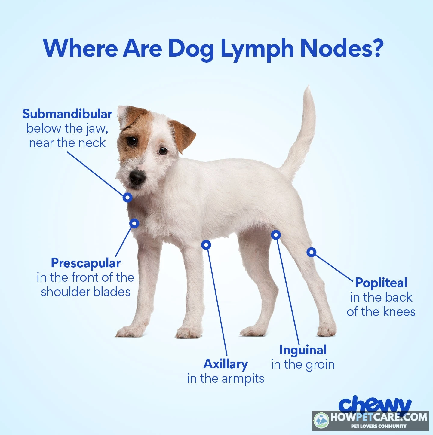
Where are lymph nodes in dogs, and how can you check them?

If you check your dog at home, go gently. Have your dog stand or lie on their side and relax. Use the flat pads of two or three fingers, not your nails or the very tips, and move the skin lightly over the tissue beneath it. Check the same spot on both sides and compare. Stop if your dog flinches, tenses, or pulls away. Don’t squeeze, repeatedly poke, or press hard on a lump.

-
1. Submandibular — under the jaw.
Slide your fingers along the underside of the jawbone toward the angle of the jaw, just in front of where the neck starts. Normal nodes feel like a small pea or flattened bean and can be hard to find. Grape-size or larger is clearly enlarged. What this area drains: teeth, mouth, and face — so dental or mouth disease is one possible cause of swelling here, among others.
-
2. Prescapular — in front of the shoulders.
Feel the hollow just ahead of the top of the shoulder blade, where the neck meets the chest. Roll the tissue gently between your fingers. A normal node is pea-to-bean size; a walnut-feeling lump is enlarged. Drains the head, neck, and front chest region.
-
3. Axillary — the armpits.
Run your hand up into the armpit of each front leg. In healthy dogs these nodes are usually too small or deep to feel — finding nothing is normal. A lump you CAN feel here deserves a vet visit. Do not dig around forcefully searching for them.
-
4. Inguinal — the groin.
Feel the fold where each back leg meets the belly. These nodes are also often not palpable when normal. Note: female dogs have mammary tissue here, and lumps in this region can be mammary rather than lymph node — another reason a vet should sort it out.
-
5. Popliteal — behind the knees.
This is the easiest node to feel in most dogs. Find the soft hollow directly behind the knee joint (stifle) of each back leg. Normal is almond- or bean-size and soft; grape-size or a firm, fixed lump is enlarged. Drains the lower hind leg — check for cuts, wounds, or infections on that leg and paw.
What "enlarged" means in practice. There’s no single size cutoff: a Great Dane’s normal node differs from a Chihuahua’s. Look for one side being clearly bigger than the other, a new lump, a lump that’s growing week to week, or one that feels fixed instead of sliding under the skin. Keep in mind that a lump in a lymph node location could actually be a salivary gland, an abscess, a cyst, or a tumor. Touch alone can’t identify it.
Can you tell a benign swollen node from cancer by feel?
Comparing what you feel can help you describe the lump and decide how quickly to act, but it can’t identify the cause. Veterinary testing, usually starting with a needle sample, is needed to distinguish causes reliably.
Lymph node patterns: Clues, overlaps, and when to act
| Feature | Possible inflammatory or infectious pattern | Possible cancer pattern | Important overlap and action |
|---|---|---|---|
| Pain when touched | Often tender or warm — the dog may flinch | Often painless (lymphoma nodes typically do not hurt) | Painless does NOT rule out infection, and painful does NOT rule out cancer. Either way, a new lump needs checking. |
| Texture | Softer, sometimes squishy if an abscess is forming | Typically firm or rubbery | Texture ranges overlap widely — "firm" alone is not a diagnosis. |
| Mobility | Usually slides a little under the skin | May feel fixed or attached to deeper tissue, especially later | A fixed lump is a red flag regardless of cause — book promptly. |
| Number of enlarged nodes | Often one node near a local problem (bad tooth → jaw node) | Classically several nodes enlarged at once | Lymphoma can start in ONE node, and infections can enlarge several. Count raises suspicion; it does not confirm. |
| Growth speed | May swell quickly over days, then plateau or shrink as the trigger resolves | Often steady enlargement over weeks | Rapid growth in either column means faster vet contact, not a home answer. |
| Other signs | Fever, lethargy, or a nearby wound, skin infection, or dental disease | Weight loss, reduced appetite, low energy — but early lymphoma dogs can seem normal | Sick-feeling dog + enlarged nodes = same-day call. Well-seeming dog + enlarged nodes = routine visit within days, not months. |

Bottom line. The table can help you describe the lump and decide how quickly to act, but it can’t tell you what it is. Veterinary testing, usually starting with a needle sample, is needed for that.
How veterinarians diagnose and treat enlarged lymph nodes
At the visit, the veterinarian will examine your dog and ask about the lump: when you first noticed it, whether your dog has been chewing on one side, any visible wounds or recent tick bites, travel history, and changes in appetite, energy, or weight. From there, the veterinarian may recommend tests based on the exam and your dog’s history.
Fine needle aspiration and cytology. If the veterinarian thinks a node should be sampled, they may start with a fine needle aspiration (FNA). A thin needle collects cells from the node. Those cells are spread on a slide and examined under a microscope (cytology). The test can often distinguish inflammatory cells, infectious agents, and cancer cells. An FNA isn’t necessary for every dog at the first visit. If the lump is small and your dog is acting completely normally, the veterinarian may recommend monitoring it first.
Bloodwork. A complete blood count (CBC) and chemistry panel can show signs of infection, provide information about organ function, and help assess overall health. Certain patterns, including changes in specific white blood cells, can point toward causes such as lymphoma.
Infectious-disease testing. Your veterinarian may recommend tests for tick-borne diseases (Lyme, Ehrlichia, Rocky Mountain spotted fever) or regional fungal diseases such as Valley fever, depending on where your dog lives and their lifestyle. These tests can help identify or rule out infection as the reason for enlarged nodes.
Imaging. X-rays or ultrasound can help the veterinarian check internal organs, look for masses elsewhere, or plan a biopsy. Ultrasound may also show more detail about the lymph nodes themselves.

Biopsy. If early test results don’t give a clear answer, or the veterinarian needs a tissue diagnosis, they may perform a surgical biopsy. This provides a larger sample of lymph node tissue for histopathology.
Treatment connected to diagnosis. Treatment depends on the cause. Antibiotics are appropriate for a bacterial infection, but they won’t help with every cause of swollen lymph nodes and won’t affect lymphoma. Dental or wound care may be needed if a local infection is responsible. Immune-mediated disease may call for targeted immunosuppressive medication. For lymphoma, oncology options include chemotherapy protocols and, in some cases, radiation or immunotherapy.
Prognosis by cause. Reactive swelling may go down as the underlying problem is treated. For example, after treatment for a dental infection, the nodes may gradually shrink. Outcomes for infections, immune-mediated disease, and cancer depend on the diagnosis and how the dog responds. Finding the cause sooner tends to leave more options and improve the outlook.
If it is lymphoma: Remission and survival expectations
For common multicentric lymphoma, these qualified ranges come from mainstream veterinary consensus:
- Without treatment. Survival is often around 4–6 weeks from diagnosis. The timeline varies with the dog’s overall health, lymphoma subtype, and individual response.
- Multi-agent chemotherapy (e.g., CHOP protocol, approximately 6 months). Many dogs go into remission with multi-agent chemotherapy. Typical median survival time is around 10–14 months. Remission means the cancer is undetectable or substantially reduced, not cured; relapse is common.
- Single-agent or lower-intensity protocols. Some dogs receive less aggressive chemotherapy. Remissions may be shorter (often 3–6 months), but side effects and costs may be lower. Discuss the trade-off between expected remission length and treatment intensity with the veterinarian.
- Factors that influence outcome. Subtype (B-cell vs. T-cell), stage at diagnosis, chemotherapy protocol, and the dog’s individual response all affect survival time. Your veterinarian can give a more personalized outlook based on your dog’s case.

What diagnosis and treatment may cost in the United States (2026)
The figures below are typical ballpark ranges drawn from US veterinary fee surveys and clinic-published prices (2023–2024). Your bill will depend on your region, clinic type, and dog’s size. Ask for a written estimate before agreeing to tests.
Estimated US costs for diagnosis and treatment
| Test or treatment | Typical estimated cost | Per test, visit, or course | Possible additional charges |
|---|---|---|---|
| Office exam (general practice) | $50–$100 | Per visit | Emergency clinic exam: $150–$250 |
| Fine needle aspiration (FNA) + cytology | $100–$300 | Per node sampled | Pathologist interpretation fee, sampling a second node |
| Bloodwork (CBC + chemistry panel) | $100–$250 | Per test | Add-on thyroid or specialty panels |
| Tick-borne / infectious disease panel | $100–$250 | Per test | Individual tests vs. bundled panels vary in price |
| X-rays (2–3 views) | $150–$400 | Per study | Sedation if the dog won’t stay still |
| Ultrasound | $300–$600 | Per study | Specialist (radiologist) review fee |
| Surgical biopsy + histopathology | $500–$1,500+ | Per procedure | Anesthesia, pain meds, recheck visits — ask what is bundled |
| Straightforward infection treatment (exam + antibiotics) | $100–$300 | Per course | Recheck exam, culture testing if it doesn’t respond |
| Dental cleaning with extractions | $500–$1,500+ | Per procedure | X-rays, extractions priced per tooth |
| Lymphoma: prednisone-only palliative plan | $50–$150 | Per month | Recheck exams every few weeks |
| Lymphoma: full multi-drug chemo protocol (e.g., CHOP, ~6 months) | $4,000–$10,000+ | Full course | Staging tests, bloodwork before each dose, treating side effects |

Money-saving moves that are safe to ask about. Ask whether testing can be staged, starting with an exam and FNA before imaging. You can also request an itemized estimate that separates per-test and per-visit charges, compare referral with in-house care, check pet insurance coverage if you enrolled before the diagnosis, or ask about nonprofit funds and payment plans for cancer care. Chemo costs vary most by protocol. A single-drug plan can cost a fraction of CHOP, with a shorter expected remission; discuss that trade-off with your veterinarian.
Your 24–72-hour monitoring and vet-visit checklist
Contact your veterinarian when you find a new lump. If your dog otherwise seems well, arrange a visit within a few days. The 24–72-hour period is for monitoring while you await advice or an appointment, not a reason to delay care if your dog worsens.
Record keeping. Note the date, location, and approximate length and width; use a ruler if possible. Take a photo with a scale, and track changes in other node areas, appetite, energy, and signs of pain. Check gently and only as needed. Use the copy-paste lump tracking log later in this article.

Stop waiting and seek same-day veterinary guidance if the lump grows quickly, new areas become enlarged, or your dog develops pain, fever, lethargy, or refuses food. For breathing or swallowing difficulty, collapse, or rapid deterioration, go to an emergency clinic immediately.
Do not squeeze, massage, lance, or apply home remedies to the lump. Don’t give human pain medicines or leftover antibiotics. Bring your dog’s exposure history, medication list, and photos to the appointment.
Copy-paste lump tracking log (for the wait before your appointment)
Fill in one entry each day while you wait for the appointment. Bring the completed log and your photos to the vet. In each photo, place a ruler or coin beside the lump as a fixed reference. That makes day-to-day growth easier to spot than relying on memory.
DOG LUMP TRACKING LOG
Date/time: ____________
Lump location (e.g., "left side under jaw"): ____________
Size — measured with ruler: ____ mm long x ____ mm wide
  (comparison object: pea / bean / grape / walnut)
Feels: soft / firm / rubbery | moves under skin? yes / no

Pain when gently touched? none / mild / dog pulls away
New lumps found in other areas? yes — where: ____________ / no
Photo taken with coin or ruler next to lump? yes / no
Appetite today: normal / reduced / refused food
Energy today: normal / quieter than usual / very lethargic
Other changes (cough, vomiting, drinking more, limping): ____________

ESCALATION CHECK — if ANY box is ticked, call the vet today
or go to an emergency clinic:
[ ] Lump visibly bigger than yesterday
[ ] New swollen areas since last check
[ ] Lump now painful
[ ] Lethargy, fever-feeling ears/body, or refused food
[ ] Any trouble breathing or swallowing → EMERGENCY NOW
Rules while monitoring. Check gently once a day at most. Repeated handling can irritate the tissue and make your size notes less reliable. Don’t squeeze, massage, lance, or apply home remedies. Human painkillers (ibuprofen and naproxen are dangerous for dogs) and leftover antibiotics can cause problems; partially treating an infection can also confuse test results. For a stable, single lump in a well dog, limit monitoring to the few days before a routine appointment, not weeks. If the appointment is more than a week away, call and ask for an earlier slot or a place on the cancellation list.
Check gently once a day at most. Never squeeze, massage, lance, or apply home remedies to the lump. Do not give human painkillers or leftover antibiotics. If the veterinarian appointment is more than a week away, call back to ask about sooner availability or a cancellation list.
Final next steps
Swollen lymph nodes in dogs can have many causes, from a local infection to serious illness affecting the whole body. Carefully noting where the lump is, how it feels, whether other areas are swollen, and how your dog is acting can help you explain what you’ve observed and decide how urgently to seek care. If you’ve found a new lump, contact your veterinarian to discuss what to do next. Depending on your dog’s condition, that may mean monitoring for a few days, booking a routine appointment within the next several days, or seeking same-day assessment. Bring your tracking log, photos, and notes about recent health, behavior, and possible exposures. Your veterinarian can determine the cause and recommend a treatment plan for your dog.

| Lymph Node Name | Body Location | Normal Size Feeling | Enlarged Size Feeling | Body Region Drained | Source |
|---|---|---|---|---|---|
| Submandibular (Mandibular) | Under the jaw (underside of the jawbone toward the angle of the jaw, just in front of where the neck starts) | Small, pliable, pea-to-flattened-bean size; can be hard to find | Grape-to-golf-ball size or larger; firm and swollen | Head, mouth, teeth, tongue, face, and snout | [1], Inferred |
| Prescapular | In front of the shoulders (hollow just ahead of the top of the shoulder blade, where the neck meets the chest / lower neck) | Small, flat, soft, pea-to-bean size; slides easily under the skin | Walnut-size or larger prominent, firm bulge | Head, neck, shoulders, front chest region, and front legs | [1], Inferred |
| Axillary | Armpit (axillary region, deep within the armpit of each front leg) | Very small, soft; usually too small or deep to feel (non-palpable in normal dogs) | Easily felt nodule or lump | Chest wall, chest cavity, and upper front legs | [1], Inferred |
| Inguinal | Groin area (inner fold where each back leg meets the belly / lower abdomen) | Small, soft; often not palpable / difficult to feel under normal tissue | Swollen, firm lump or structure (note: may also be mammary tissue in females) | Hind legs, lower abdomen, vulva/penis, and mammary glands | [1], Inferred |
| Popliteal | Behind the knees (soft hollow directly behind the knee joint/stifle of each back leg) | Small, soft, moveable, almond- to pea- or bean-size node tucked behind the joint | Grape-size or larger prominent, firm mass or fixed lump | Lower hind legs, feet/paws, and tail area | [1], Inferred |
FAQ
How long does lymph node swelling last in dogs?
It depends on the cause: a reactive node may shrink after the underlying problem is treated, while other causes persist or progress. Ask your veterinarian when to expect improvement and arrange a recheck if the swelling persists or worsens.
Can a dog’s lymph nodes swell after vaccination?
A nearby lymph node can sometimes enlarge temporarily after vaccination as part of an immune response. Tell your veterinarian about the vaccine and any new lump, especially if it grows, persists, or your dog seems unwell.
Can swollen lymph nodes in dogs go away on their own?
Some reactive swelling can subside when a minor trigger resolves, but a lump cannot be safely identified or diagnosed by waiting for it to disappear. Contact your veterinarian about a newly noticed lump and follow the recommended examination timeline.
Are swollen lymph nodes in dogs contagious to people or other pets?
Swollen nodes themselves are not contagious, but an underlying infection sometimes can be. Tell your veterinarian about exposures and follow their hygiene and separation advice until the cause is clearer.