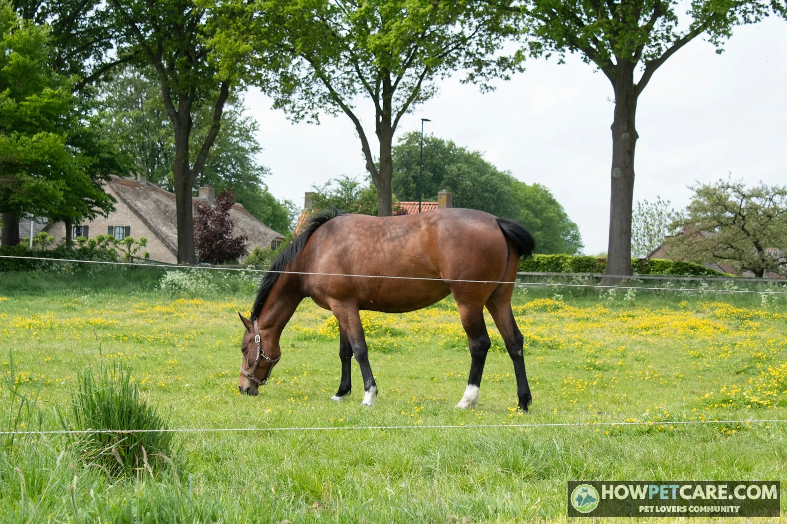

Every horse is described using the same core regions and landmarks:
- Head and neck: poll, forelock, muzzle, crest, throatlatch
- Body and topline: withers, back, loin, barrel, flank, croup, dock
- Foreleg (top to bottom): shoulder, elbow, forearm, knee (carpus), cannon, fetlock, pastern, hoof
- Hind leg (top to bottom): hip, thigh, stifle, gaskin, hock (tarsus), cannon, fetlock, pastern, hoof
- Hoof: wall, sole, frog, bars, white line, coronary band; inside, the laminae, coffin bone, navicular bone, and digital cushion
A horse’s major body regions are the head and neck, body, forequarters, hindquarters, legs, and hooves. Within them, terms such as poll, withers, fetlock, and hock point to specific landmarks used in lessons, veterinary conversations, and conformation discussions. The labeled maps and glossary below follow the horse from nose to tail, then down each leg to the hoof.
Parts of a Horse, Labeled: External Points From Nose to Tail
Use this visual map to follow the body from head to tail, then trace each limb toward the ground. These external landmarks are often called the points of the horse.
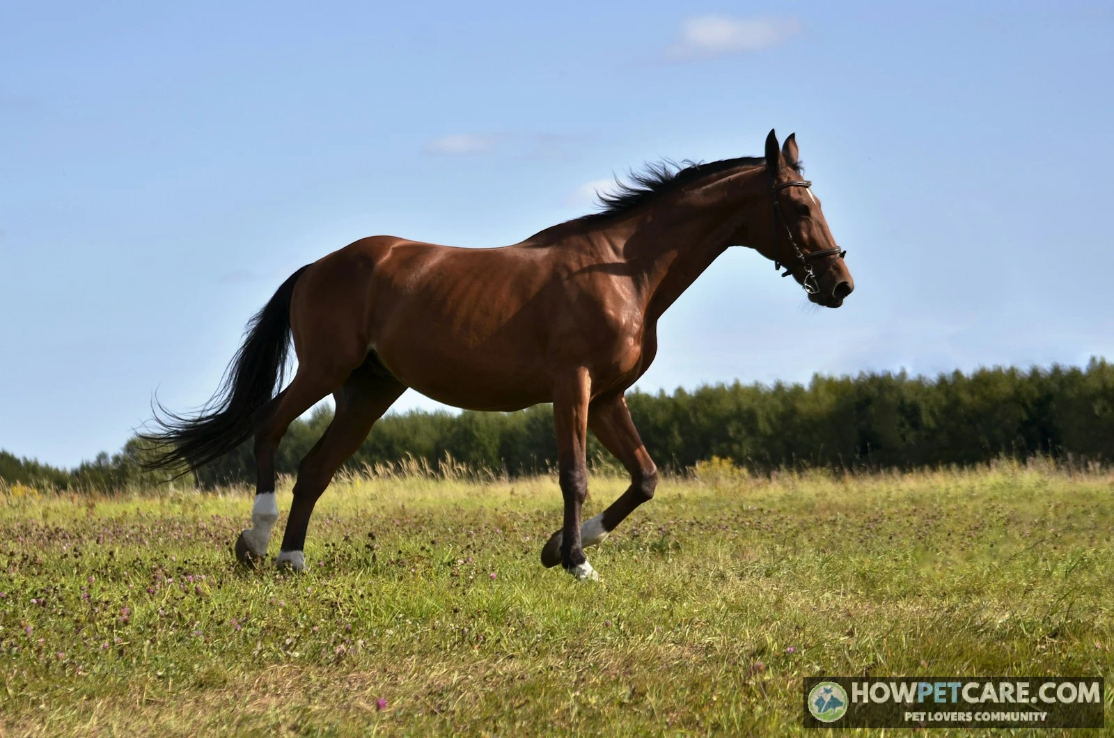
Diagram 1: External points, viewed from the side
HEAD AND NECK                         BODY AND HINDQUARTERS

 ears → poll → crest → withers → back → loin → croup → dock → tail
   |           neck       |                |       |

forelock        |      shoulder          barrel   hip

   |        throatlatch   |                |       |

 eyes                     chest          flank    thigh
   |

 muzzle

FORELIMB                              HIND LIMB
 shoulder                             hip and thigh
    ↓                                      ↓
 elbow                                  stifle
    ↓                                      ↓
 forearm                                gaskin
    ↓                                      ↓
 knee                                   hock
    ↓                                      ↓
 cannon                                 cannon
    ↓                                      ↓
 fetlock                                fetlock
    ↓                                      ↓
 pastern                                pastern
    ↓                                      ↓
 hoof                                   hoof
This map shows locations, not scale. The limb paths are simplified; the definitions below place each landmark more precisely.
Head and neck: poll, forelock, muzzle, crest, throatlatch
- Poll: The area at the top of the head between and just behind the ears, near where the skull meets the neck.
- Forelock: The hair that falls forward between the ears and over the forehead.
- Muzzle: The soft nose and mouth area at the front of the face, including the nostrils and lips.
- Crest: The upper edge of the neck, where the mane grows.
- Throatlatch: The area under the jaw where the head meets the neck. The word can also refer to a bridle strap.
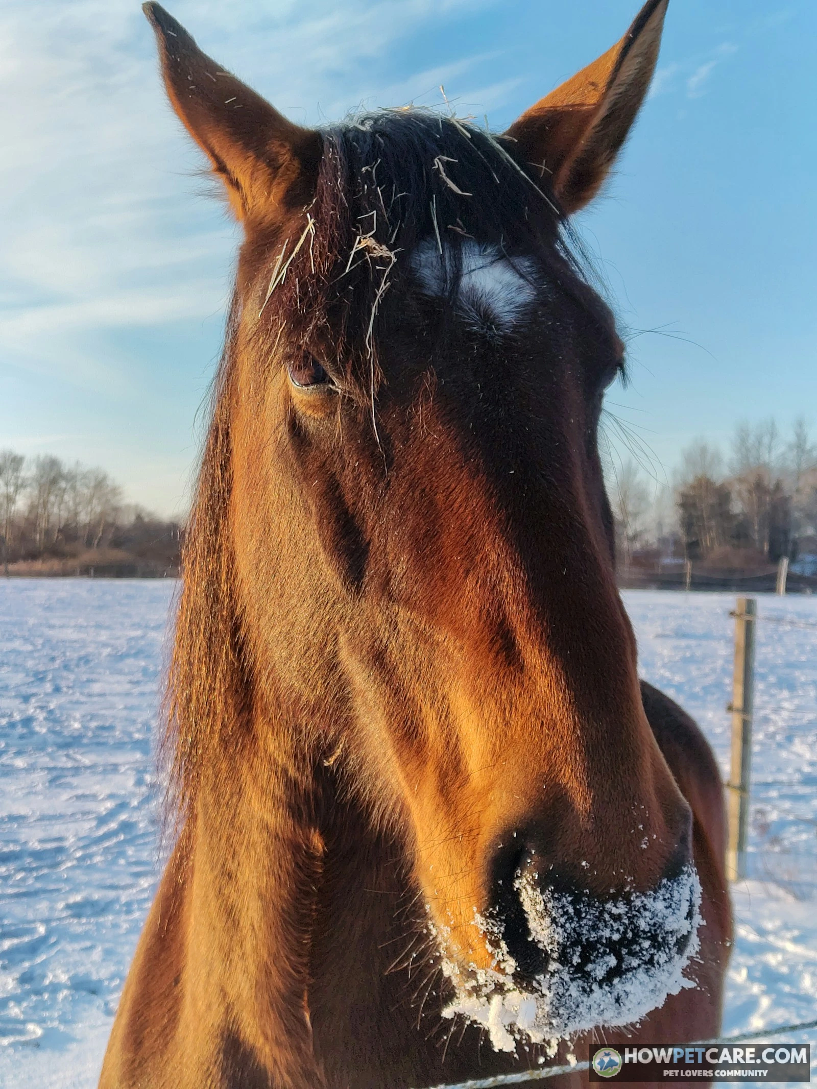
Body and topline: withers, back, loin, barrel, croup, dock
- Withers: The raised ridge where the neck meets the back, formed largely by the upward projections of thoracic vertebrae. Height is measured here, and the withers are also an important saddle-fit reference.
- Back: The upper body behind the withers and ahead of the loin.
- Loin: The short upper section between the last rib and the croup, over the lumbar region.
- Barrel: The rounded middle of the body, including the rib-cage and belly regions.
- Flank: The area behind the ribs and ahead of the hind leg, below the loin.
- Croup: The upper contour of the hindquarters, from the loin toward the tail head.
- Dock: The fleshy base of the tail, beneath the long tail hair.
Forequarters vs hindquarters: what each term includes
The forequarters comprise the shoulders, chest, and forelimbs. The hindquarters include the hip, croup, thighs, and hind limbs. In general, the forequarters carry more of the horse’s weight, while the hindquarters provide much of its forward drive.
To check your recall, cover Diagram 1 and point to the withers, barrel, croup, and poll on a horse picture. Then uncover the map and check their order.
Horse Leg Anatomy and Terminology: Foreleg and Hind Leg, Joint by Joint
The foreleg’s knee is anatomically the carpus, or wrist region. The hind leg’s stifle corresponds to the human knee, while the hock corresponds to the ankle region. Learn each limb from the body downward toward all four hooves; the same lower-leg terms apply to all four limbs.
Diagram 2: Limb landmarks and underlying structures
FORELIMB                              HIND LIMB
Shoulder: scapula and shoulder joint  Hip: pelvis and hip joint
              ↓                                   ↓
Elbow: humerus meets radius           Thigh: femur region
              ↓                                   ↓
Forearm: radius                       Stifle: femur, tibia, patella
              ↓                                   ↓
Knee: carpus                          Gaskin: tibia region
              ↓                                   ↓
Cannon: third metacarpal              Hock: tarsus
              ↓                                   ↓
Fetlock: metacarpophalangeal joint    Cannon: third metatarsal
              ↓                                   ↓
Pastern: phalangeal region            Fetlock: metatarsophalangeal joint
              ↓                                   ↓
Coronet → hoof                        Pastern → coronet → hoof
Foreleg: shoulder to hoof
- Shoulder: The sloping region behind the neck, containing the shoulder blade, or scapula, and shoulder joint.
- Elbow: The joint where the upper foreleg meets the forearm, close against the chest.
- Forearm: The region between elbow and knee, supported mainly by the radius.
- Knee: The prominent joint region between forearm and cannon. Its formal name is the carpus (KAR-pus).
- Cannon: The long, relatively straight region below the knee. Its main bone is the third metacarpal.
- Fetlock: The joint below the cannon and above the pastern, pronounced FET-lok.
- Pastern: The sloping region from fetlock toward the hoof, pronounced PAS-tern.
- Coronet: The band where the hairline meets the hoof wall, also called the coronary band.
- Hoof: The tough outer capsule that encloses and protects the end of the digit.
Hind leg: hip, stifle, gaskin, and hock
- Hip: The upper hindquarter region around the pelvis and hip joint.
- Thigh: The muscular region between hip and stifle, over the femur.
- Stifle: The joint region high on the hind limb, near the lower flank, pronounced STY-ful. It includes the femorotibial and femoropatellar joints.
- Gaskin: The muscular region between stifle and hock, over the tibia; pronounced GAS-kin.
- Hock: The joint region between gaskin and hind cannon, formally the tarsus. Its backward-projecting point is the heel bone.
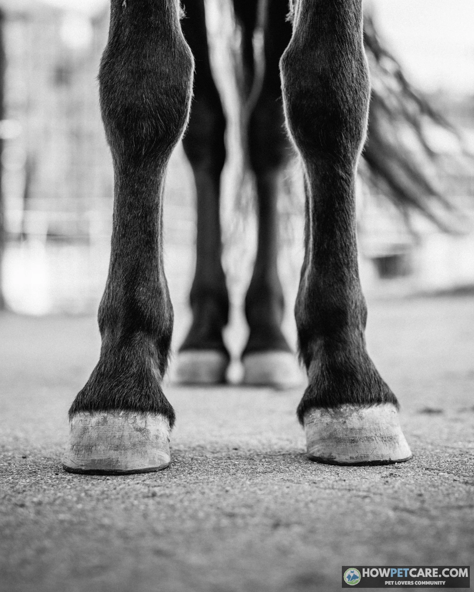
Cannon bone, fetlock, and pastern: the lower-limb segments beginners mix up
The cannon is the long segment above the fetlock, which is the joint at its lower end. Below that joint, the pastern slopes toward the hoof. The main cannon bone is the third metacarpal in front and the third metatarsal behind; the corresponding fetlock joint names also differ by limb.
 You might be wonderingHorse blanket sizesHorse Blanket Sizes: Printable 36–90 Inch Chart & How to MeasureRead the answer
Hands, Height, Ages, and Sexes: Measuring and Naming Horses Correctly
How to measure a horse in hands
A hand is four inches. Measure height vertically from level ground to the highest point of the withers, not to the ears or top of the head.
Stand the horse squarely on level ground, with its head in a natural position. Use a measuring stick designed for horses, or a suitable tape with a level reference across the withers. An experienced handler can help keep the horse still.
The number after the point in a hands measurement gives additional inches, not a decimal fraction of a hand:
- 14.2 hands: 14 × 4 + 2 = 58 inches, or 147.3 centimeters rounded.
- 15.3 hands: 15 × 4 + 3 = 63 inches, or 160.0 centimeters rounded.
- 16.0 hands: 16 × 4 = 64 inches, or 162.6 centimeters rounded.
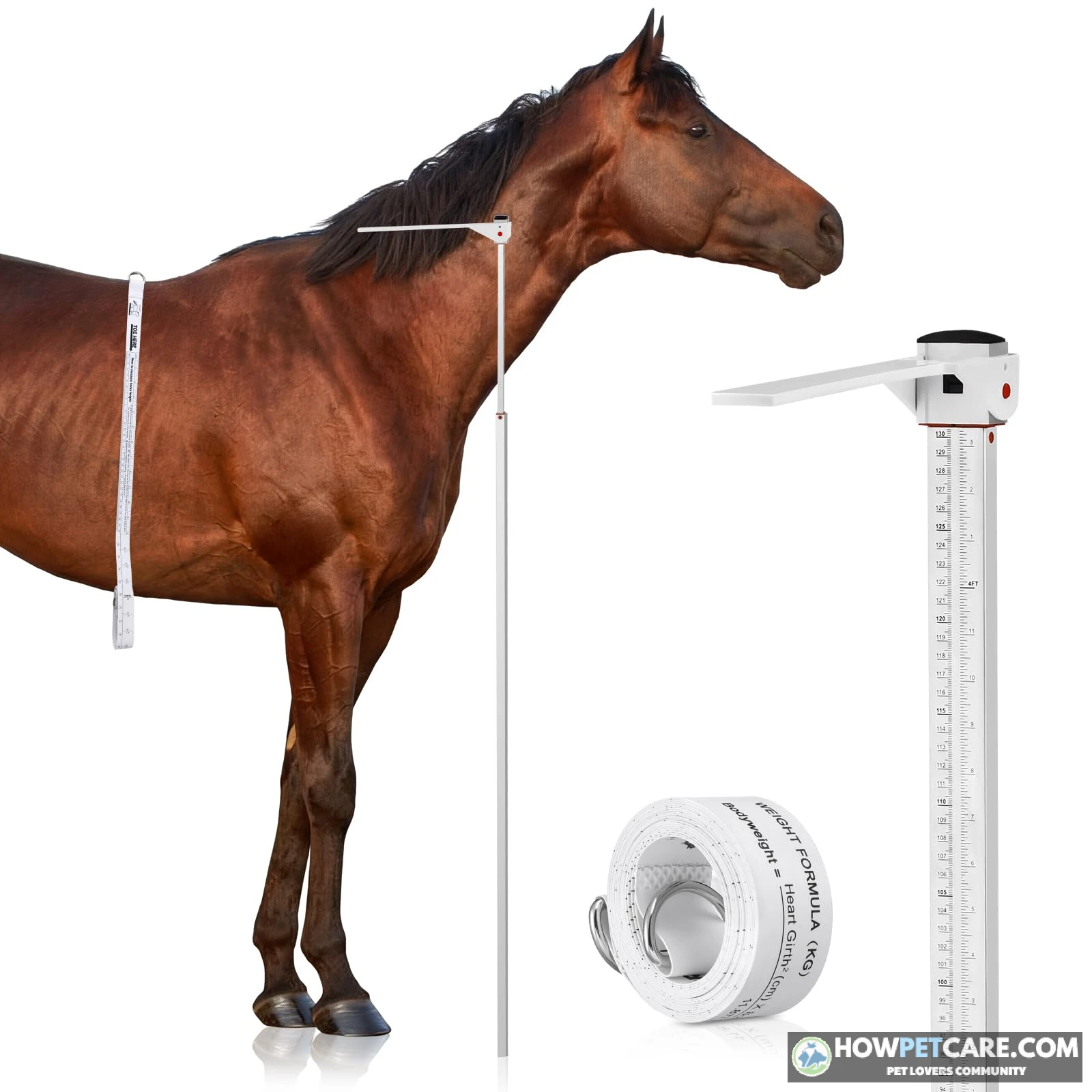
After 15.3 hands comes 16.0, not 15.4. The notation 14.2 means 14 hands plus 2 inches, not 14.2 multiplied by 4.
Horse vs pony: where the line falls and why it is not absolute
Under the usual height convention, an equine measuring 14.2 hands or less is a pony; one over 14.2 hands is a horse. Breed identity and competition rules can create exceptions. Miniature Horses, for example, are called horses despite their height. When classification matters, check the relevant organization’s rules.
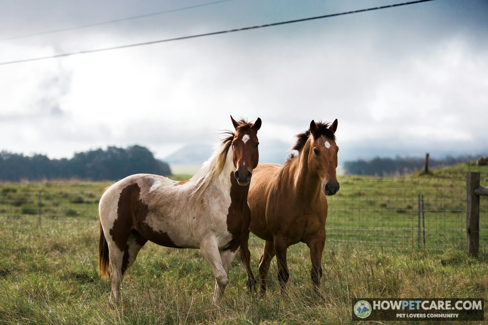
Foal, colt, filly, mare, stallion, gelding: age and sex terms side by side
| Term | Sex | Typical age or status | Notes |
|---|---|---|---|
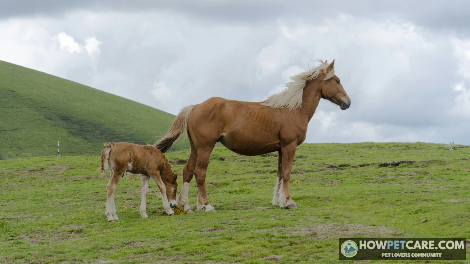
| Foal | Either | Young horse, generally under one year | A general age term, not a synonym for colt. |
| Weanling | Either | Young horse that has been weaned, usually in its first year | Describes separation from nursing. |
| Yearling | Either | One-year-old | Applies to either sex. |
| Colt | Male | Generally under four years | A young male, not every young horse. |
| Filly | Female | Generally under four years | A young female; not simply a small mare. |
| Mare | Female | Generally four years or older | An adult female. |
| Stallion | Male | Mature, uncastrated | An intact adult male. |
| Gelding | Male | Castrated, at any age | Describes status rather than a particular adult age. |
Age conventions differ among some breed registries and racing organizations. For a female horse listing, check the horse’s actual age and the rules that apply.
Inside and Outside the Hoof: A Cross-Section Guide to Hoof Anatomy
The hoof has a visible horn capsule and living structures inside it. These maps distinguish what you can see from what lies beneath in a horse’s hoof; neither is drawn to scale.
Diagram 3A: Hoof viewed from underneath
                 TOE
          __________________
         /   hoof wall
        /  ----------------
       |  white line         |

       |        sole         |

       |   bar  /  bar      |

       |       /            |

       |      /frog         |

            /             /
          heel    heel    /
          ----------------
                 BACK
Diagram 3B: Simplified side cross-section
                    FRONT / TOE
      coronary band
            ↓
 hair  ____/================  ← hoof wall
      /      laminae      /  ← between wall and underlying tissue
BACK | digital cushion   /

     | navicular bone   / ← behind coffin bone

     |      coffin bone/  ← distal phalanx

     | deep flexor tendon   ← passes behind navicular bone

      ____frog____sole____/
What you can see: wall, sole, frog, heel, and coronary band
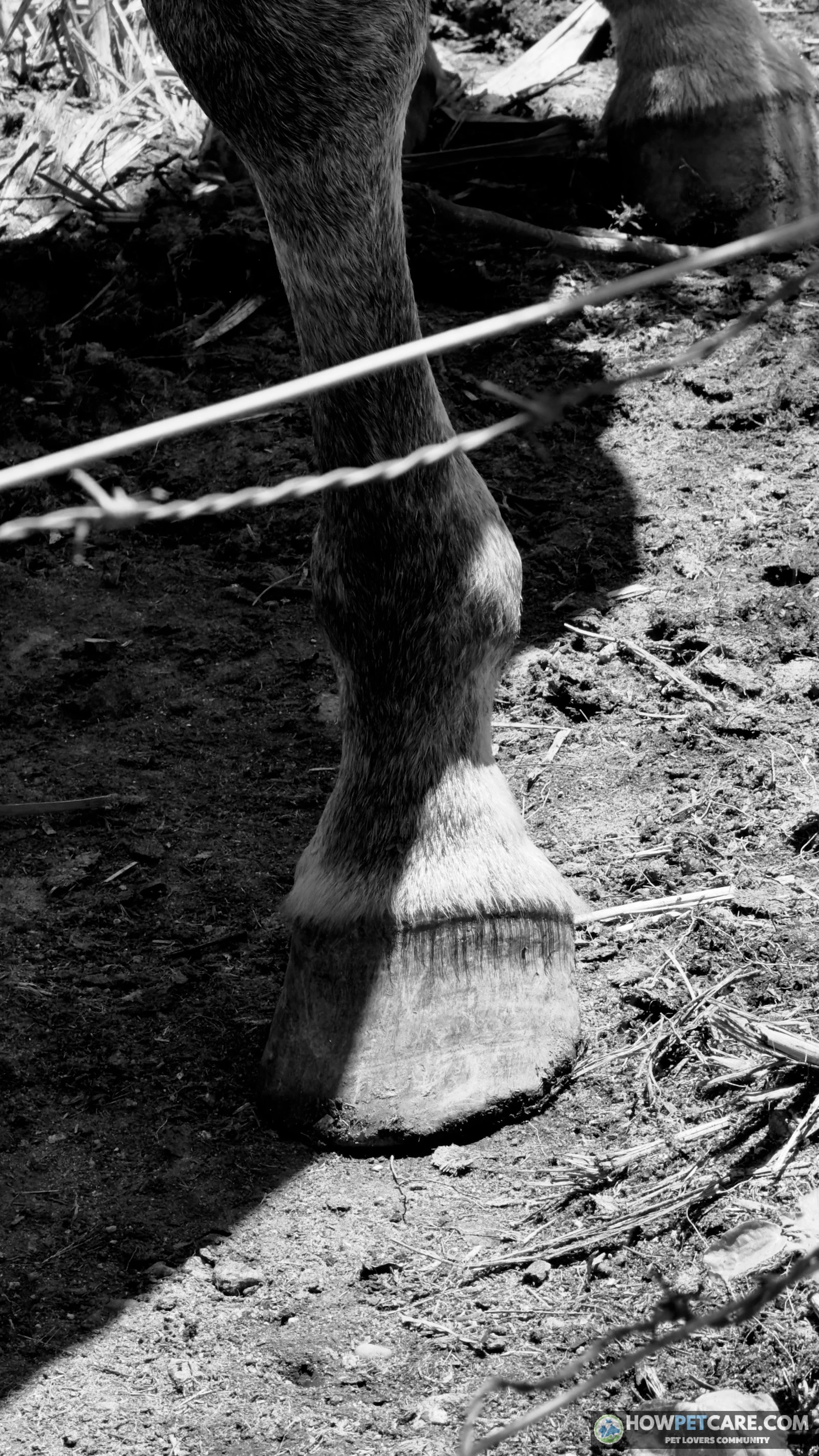
- Wall: The hard outer horn visible around the hoof, protecting the structures inside.
- Sole: The surface inside the wall on the underside, helping protect the foot.
- Frog: The V-shaped, rubbery structure on the underside, pointing toward the toe.
- Heel: The rear part of the hoof, on either side of the frog.
- Bars: Inward extensions of the wall on the underside, running forward from the heel alongside the frog.
- White line: The junction visible on the underside between the wall and the sole region.
- Coronary band: The band at the hairline from which most of the hoof wall grows.
What you cannot see: laminae, coffin bone, navicular bone, and digital cushion
- Laminae (LAM-ih-nee): Interlocking layers of sensitive and insensitive tissue that help suspend the coffin bone within the hoof capsule. Laminitis refers to inflammation and damage affecting this connection.
- Coffin bone: The bone inside the front of the hoof, also called the distal phalanx or third phalanx.
- Navicular bone (nuh-VIK-yuh-ler): A small bone behind the coffin joint, also called the distal sesamoid bone.
- Digital cushion: A resilient pad of tissue at the back of the foot, above the frog.
- Deep digital flexor tendon: A strong tendon that runs down the back of the limb, over the navicular region, and attaches to the underside of the coffin bone.
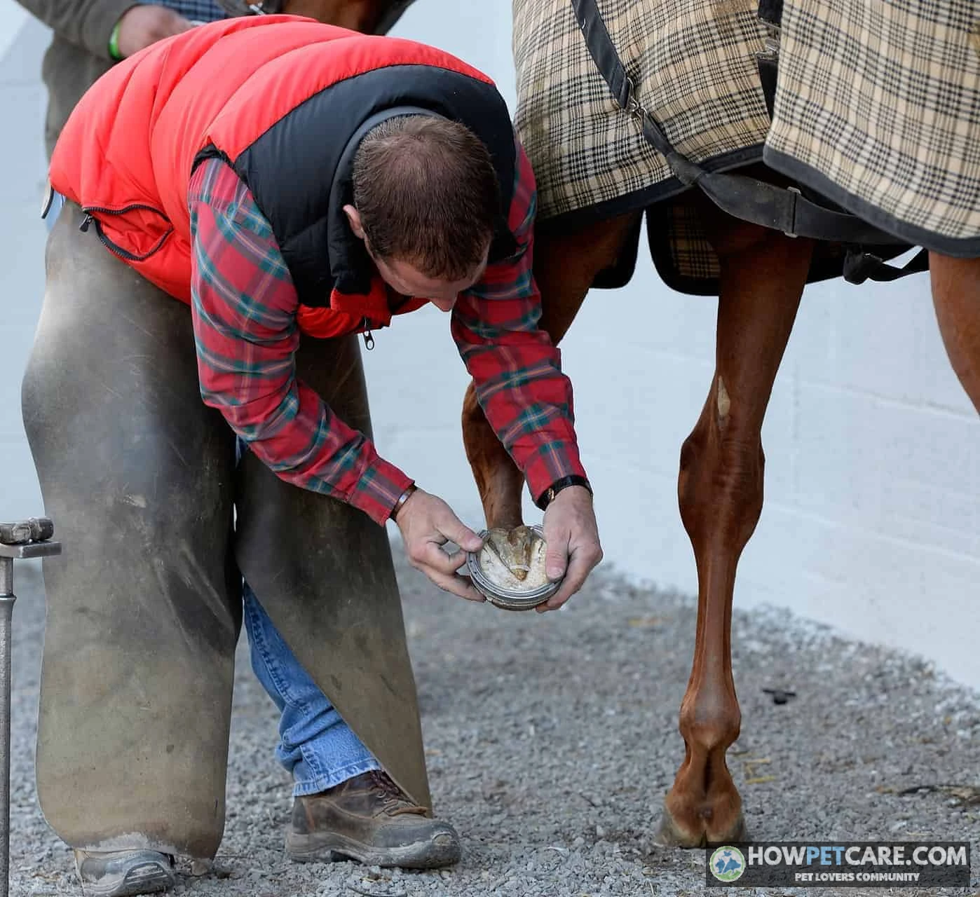
These terms help you describe a structure, not diagnose a problem. Contact a veterinarian about suspected hoof pain or disease, and involve your farrier in appropriate hoof-care decisions.
Horse Leg Joints vs Human Anatomy: One-Table Comparison
A horse bears weight on the end of one principal digit on each limb. The familiar foreleg knee corresponds to a human wrist, not a human knee. These comparisons match structures by anatomy, not by appearance or identical function.
| Horse part (barn term) | Veterinary name | Nearest human equivalent | Where to find it | Key difference |
|---|---|---|---|---|
| Shoulder | Scapula and shoulder joint | Shoulder blade and joint | Sloping region behind the neck | The horse’s forelimb connects to the trunk mainly through muscles rather than a collarbone. |
| Elbow | Elbow joint | Elbow | Against the chest, above the forearm | Similar joint, held close to the body. |
| Foreleg knee | Carpus | Wrist | Between forearm and front cannon | Called a knee in barn language, but not the anatomical knee. |
| Cannon | Third metacarpal or metatarsal | Long bone of the hand or foot | Between knee or hock and fetlock | The large central bone supports one principal digit; smaller splint bones lie alongside it. |
| Fetlock | Metacarpophalangeal or metatarsophalangeal joint | Knuckle or toe-base joint | Between cannon and pastern | The joint bears weight in a very different posture. |
| Pastern | Phalangeal region, including proximal and middle phalanges | Finger or toe segments | Sloping region below fetlock | The bones extend toward and partly into the hoof; the visible region is not identical to one bone. |
| Coffin bone | Distal phalanx | Fingertip or toe-tip bone | Inside the hoof | Enclosed within the hoof capsule. |
| Stifle | Femorotibial and femoropatellar joints | Knee | High on the hind limb | Less conspicuous against the body than the hock. |
| Gaskin | Crural region, including tibia | Lower leg or shin region | Between stifle and hock | A muscular region, not a single joint. |
| Hock | Tarsus | Ankle and heel region | Between gaskin and hind cannon | The projecting point includes the heel bone, or calcaneus. |
Below the knee and hock, the limbs consist largely of bones, tendons, ligaments, vessels, nerves, and other tissues rather than large muscle bellies. Keep this shorthand in mind: horse knee = wrist; horse hock = ankle; horse stifle = knee.
Directional Terms and Dual Vocabulary: Barn Words vs Veterinary Names
Dorsal, ventral, cranial, caudal, proximal, distal: plain-English definitions
Veterinary directions use the horse’s body as their reference, not the observer’s position.
| Term | Meaning | Example |
|---|---|---|
| Dorsal | Toward the back or upper surface; on the lower limb, toward the front surface | The dorsal hoof wall faces forward. |
| Ventral | Toward the belly or underside | The belly is ventral to the back. |
| Cranial | Toward the head; on the upper limb, toward the front | The chest is cranial to the barrel. |
| Caudal | Toward the tail; on the upper limb, toward the rear | The croup is caudal to the withers. |
| Proximal | Closer to the trunk or point of attachment | The knee is proximal to the fetlock. |
| Distal | Farther from the trunk or point of attachment | The hoof is distal to the fetlock. |
| Medial | Toward the body’s midline | The inside surface of a leg is medial. |
| Lateral | Away from the body’s midline | The outside surface of a leg is lateral. |
Below the carpus or tarsus, the rear surface is called palmar on a forelimb and plantar on a hind limb. “Distal limb” generally means a lower portion of the leg, though its exact extent depends on context.
Which terms are barn language and which are veterinary language
In lessons, knee, hock, and fetlock are practical, widely understood terms. A report may instead use carpus, tarsus, or a longer joint name. The pastern is a region containing parts of more than one phalanx, so it shouldn’t automatically be translated as just the proximal phalanx.
Both vocabularies work when speaking with a veterinarian or farrier. Use the word you know, identify the location, and ask which structure is meant if a formal term is unfamiliar. Near side means the horse’s left; off side means its right. Left and right always refer to the horse, not to you when you face it.
Using Anatomy Terms: Decoding Vet Reports, Injury Descriptions, and Conformation
Reading a vet report: region, side, and structure
Consider this fictional note: “Palpable heat and swelling of the distal right forelimb centered at the fetlock.”
- Right forelimb: The horse’s right front leg.
- Distal: Farther down that limb.
- Fetlock: The named joint around which the swelling is centered.
- Palpable: Detectable by touch.
Other report terms include lameness, an abnormality of movement; effusion, excess fluid within a joint or other enclosed structure; and laminitis, inflammation and damage involving the hoof laminae. “Distal limb swelling” gives a location and observation, not a cause.
When reporting what you see, give the side, limb, nearest landmark, and observation: “left hind, around the fetlock, swelling.” Your veterinarian can ask follow-up questions and assess what the finding means.
Conformation basics: cosmetic quirks vs traits linked to soundness
Conformation describes the arrangement and proportions of a horse’s body. Discussions may cover the topline, shoulder angle, leg alignment, pastern slope, or hoof shape. Common descriptions include:
- Straight: A description of alignment. Clarify whether it refers to a leg viewed from the front, side, or rear.
- Upright pastern: A pastern with a relatively steep slope when viewed from the side.
- Sickle hock: A hind-limb alignment in which the hock appears excessively angled when viewed from the side.
- Base-narrow: A stance in which the feet are closer together than the limbs’ points of origin higher up.
Coat color and many markings are largely cosmetic. Alignment and hoof shape can affect how forces pass through a limb and may be associated with soundness concerns, but appearance alone can’t predict lameness, disease, or performance. Ask a veterinarian or qualified equine professional to assess the individual horse.
Sale listings may describe height in hands, age and sex, conformation, and gait. A walk, trot, canter, and gallop are gait terms. In Western contexts, jog and lope commonly refer to slower versions of the trot and canter. Treat a listing’s wording as a description to clarify, not a substitute for an independent evaluation.
Horse Anatomy Study Kit: Pronunciation Glossary, Printable Chart, and Self-Quiz
Glossary by region with pronunciation
Print this chart as a compact equine anatomy reference. Formal names may identify a specific bone or joint within a broader barn-language region rather than serve as exact synonyms.
| Body region | Lay or barn term | Pronunciation | Veterinary or formal term | Location cue |
|---|---|---|---|---|
| Head | Poll | POHL | Poll or occipital region | Top of head between and just behind ears. |
| Head | Muzzle | MUZ-ul | Muzzle or rostral facial region | Nose and mouth. |
| Neck | Crest | KREST | Dorsal neck | Upper edge of neck beneath mane. |
| Neck | Throatlatch | THROHT-lach | Throatlatch region | Under jaw where head meets neck. |
| Body | Withers | WITH-erz | Region of thoracic spinous processes | Ridge where neck meets back. |
| Body | Barrel | BAIR-ul | Thoracic and abdominal regions | Rounded middle of body. |
| Body | Loin | LOYN | Lumbar region | Between last rib and croup. |
| Body | Croup | KROOP | Rump or sacral region | Top of hindquarters toward tail head. |
| Body | Dock | DOK | Tail or caudal base | Fleshy part of tail. |
| Foreleg | Knee | NEE | Carpus, KAR-pus | Between forearm and front cannon. |
| Hind leg | Stifle | STY-ful | Femorotibial and femoropatellar joints | High on hind limb near flank. |
| Hind leg | Gaskin | GAS-kin | Crural region | Muscular region between stifle and hock. |
| Hind leg | Hock | HOK | Tarsus, TAR-sus | Between gaskin and hind cannon. |
| Lower limb | Cannon | KAN-un | Third metacarpal or metatarsal | Long region above fetlock. |
| Lower limb | Fetlock | FET-lok | Metacarpophalangeal or metatarsophalangeal joint | Joint below cannon. |
| Lower limb | Pastern | PAS-tern | Phalangeal region | Sloping region below fetlock. |
| Hoof | Coronet | KOR-uh-net | Coronary band | Hairline at top of hoof wall. |
| Hoof | Laminae | LAM-ih-nee | Dermal and epidermal laminae | Interlocking tissues inside hoof wall. |
| Hoof | Navicular bone | nuh-VIK-yuh-ler | Distal sesamoid bone | Behind the coffin joint inside the hoof. |
Label-it-yourself diagram exercise
Print the labeled maps and this blank chart using your browser’s print function. Fill in the blanks from memory, then check against Diagrams 1–3. For another round, trace a horse outline and add the same landmarks.
TOPLINE:  ears → _____ → crest → _____ → back → loin → _____ → dock
FORELEG:  shoulder → elbow → forearm → _____ → cannon → _____ → pastern → hoof
HIND LEG: hip → thigh → _____ → gaskin → _____ → cannon → fetlock → hoof
HOOF:     outer _____; underside _____ and frog; inside _____ bone and laminae
The missing terms, in order, are poll, withers, croup; knee, fetlock; stifle, hock; wall, sole, coffin.
Ten-question self-quiz
Cover the answers and try these on paper or with a study partner:
- Which ridge is the reference point for height?
- What human joint corresponds to the horse’s foreleg knee?
- Which hind-leg joint corresponds to the human knee, and which to the ankle?
- What are the three landmarks between knee or hock and hoof, in order?
- How many inches is 15.2 hands?
- What does “distal left forelimb” identify?
- What tissues help connect the hoof wall to the structures supporting the coffin bone?
- What is the usual height boundary for a pony?
- What are a young female and a castrated male called?
- What does dorsal mean on the body and on the front of the lower limb?
Answers: 1. Withers. 2. Wrist. 3. Stifle and hock, respectively. 4. Cannon, fetlock, pastern. 5. 62 inches. 6. A lower or farther-from-the-body portion of the left front leg. 7. Laminae. 8. 14.2 hands, with classification exceptions. 9. Filly and gelding. 10. Toward the back or upper surface on the body; toward the front surface on the lower limb.
Put the Terms to Work: A Head-to-Hoof Observation Routine and Next Steps
A brief, consistent observation routine helps you practice the vocabulary and notice changes worth reporting. Stay in a safe position, and ask an experienced handler for help if you don’t know the horse.
- Look at the eyes, face, and muzzle, then follow the head to the poll and neck.
- Check the outline of the withers, back, and loin, followed by the barrel and flank.
- Turn your attention to the croup and dock.
- Observe each leg from shoulder or hip down through the named joints to the hoof. Compare left and right where practical.
- Record what you notice with its side and location, such as “left hind, fetlock, swelling,” and share it with the horse’s caretaker or veterinarian.
Call a veterinarian about new heat, swelling, lameness, or a concerning change in appetite or behavior. Don’t try to diagnose it from terminology alone. To continue studying, learn the external points first, then the limbs and hoof, followed by directional and veterinary terms. Separate hoof-care, weight-estimation, and mare-care resources can offer more detail when those topics become relevant.
 You might be wonderingHolsteiner horseHolsteiner Horse Guide: Traits, Costs, Bloodlines & FitRead the answer
FAQ
What are normal resting temperature, pulse, and respiration for an adult horse?
An adult horse at rest typically has a temperature of about 99 to 101.5°F (37.2 to 38.6°C), a pulse of about 28 to 44 beats per minute, and a respiratory rate of about 8 to 16 breaths per minute. Readings outside these ranges, or a change from your horse’s own normal, are worth discussing with a veterinarian.
What are chestnuts and ergots on a horse?
Chestnuts are flat, horny growths on the inner side of each leg, above the knee on the front legs and below the hock on the hind legs. Ergots are small horny nubs hidden in the tuft of hair at the back of each fetlock. Both are normal.
What do ‘near side’ and ‘off side’ mean?
The near side is the horse’s left side and the off side is the right. Handlers traditionally lead, mount, and groom from the near side, so the terms often appear in riding instruction.
How many bones does a horse have?
A horse has about 205 bones. The exact count varies slightly between individuals and breeds, mainly because of differences in the number of vertebrae.
Is the horse’s knee the same as a human knee?
No. The joint called the knee on a horse’s foreleg corresponds to the human wrist (the carpus). The joint that corresponds to the human knee is the stifle on the hind leg.