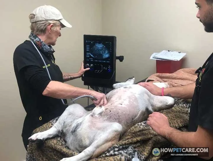

A dog ultrasound is a non-invasive imaging scan that uses sound waves—not radiation—to examine soft tissues, organs, blood flow, or pregnancy in real time. A typical scan takes 20–60 minutes, usually without sedation, and costs about $300–$600 in the US before add-ons like sampling or specialist review.
A dog ultrasound uses high-frequency sound waves, rather than ionizing radiation, to examine soft tissues, organs, blood flow, or pregnancy in real time. A typical scan takes about 20–60 minutes, and many dogs don’t need sedation. The right test, total bill, and preparation depend on what your veterinarian needs to see and whether sampling is planned.
Before approving the appointment, ask what the scan is intended to answer and whether the estimate includes the exam and image interpretation. Check which charges might be added for sedation, sampling, laboratory work, or specialist review. Ultrasound can reveal abnormalities, but an image alone can’t establish whether a mass is cancerous. Here’s what the appointment involves, how to prepare, what the costs may add up to, and how to use the report to plan what happens next.
Dog ultrasound: what to expect and when it helps
A veterinary ultrasound, also called a sonogram, creates moving images from echoes made when sound waves meet tissues inside the body. Unlike an X-ray, it doesn’t use ionizing radiation. It also lets the veterinarian assess some organs and structures in real time.
A typical appointment includes check-in, preparation, scanning, and a discussion of what the team observed. Staff position your dog so they can reach the area being examined. They may clip a small patch of fur, apply gel, and move a handheld probe over the skin. Most scans take about 20–60 minutes, though the appointment can take longer if the clinic needs extra preparation, a specialist review, or a sample.

A veterinarian may recommend an abdominal scan after unexplained vomiting, diarrhea, weight loss, abdominal pain or swelling, abnormal bloodwork, blood in the urine, or concern about internal bleeding, a mass, or pregnancy. Ultrasound can also help investigate heart disease or guide a sample from an abnormal area. These signs don’t automatically mean your dog needs an ultrasound. The veterinarian chooses imaging based on the examination and the question that needs answering.
If your dog is suddenly weak, collapses, has trouble breathing, has a swollen painful abdomen, or is bleeding significantly, contact a veterinarian or emergency hospital promptly instead of waiting for a routine imaging appointment. A scan may be part of an urgent assessment, but it isn’t a substitute for one.
What a dog ultrasound can show—and what it cannot
An abdominal ultrasound can help examine organs such as the liver, spleen, kidneys, and bladder. It may show changes in organ size or texture, some masses, fluid outside organs, and certain bladder stones. It can also help a veterinarian choose where to collect fluid or tissue. What the scan shows depends on the target, the dog, the equipment, and the person performing it.
Ultrasound can’t provide a complete view through every part of the body. Sound doesn’t pass well through air or bone, so gas in the digestive tract or ribs over an area can obscure what lies behind them. Image quality also depends partly on probe selection and the operator’s skill. Doppler ultrasound adds information about blood flow, but it doesn’t make every structure visible or turn an imaging appearance into a definitive diagnosis.

Abdominal, cardiac and emergency scans
An abdominal ultrasound examines organs in the abdomen. It can investigate a range of concerns, but it doesn’t necessarily evaluate every organ or answer every question in the same way. Ask which areas the veterinarian plans to scan and whether the study is comprehensive or focused.
An echocardiogram is a focused ultrasound examination of the heart. It assesses heart structure and movement, and Doppler may be used to assess blood flow. Although both use ultrasound technology, an echocardiogram is different from a routine abdominal ultrasound.
A FAST scan—short for focused assessment with sonography for trauma—is a rapid, focused examination used in emergency settings to look for fluid in selected areas. It can help guide urgent decisions, but it isn’t the same as a full abdominal study and doesn’t rule out every injury or disease.

Can a sonogram tell whether a mass is cancerous?
No. Ultrasound can describe a mass’s location, size, shape, and appearance, and it may help a veterinarian judge how concerning it looks. It can’t definitively distinguish a benign mass from a malignant one. Cytology from a fine-needle aspirate or histopathology from a tissue biopsy may be needed for a definitive diagnosis; sometimes monitoring or other tests are reasonable instead. A biopsy isn’t inevitable. The choice depends on the suspected condition, the risks of sampling, and whether the result would change treatment.

Dog ultrasound cost: build an all-in estimate
The cost of a dog ultrasound depends on the body area, clinic, region, urgency, and whether the scan is performed in general practice or at a referral center. The following are typical US estimates for 2026, not a guaranteed quote. Some clinics bundle the examination or interpretation into the scan price; others list each charge separately. Ask for a written estimate that identifies required charges and conditional add-ons.
Base scan and possible add-on charges
| Charge | Typical US price range | When it may apply | Included or separate? | Example amount |
|---|---|---|---|---|
| Office or exam fee | $50–$100 | Almost always; may be bundled | Sometimes bundled | $75 |
| Abdominal ultrasound (base scan) | $300–$600 | Core charge; emergency or specialist scans may cost more | Base fee | $450 |
| Sedation | $50–$200 | Anxious, painful, or difficult-to-position dogs; often needed for sampling | Usually separate | $120 |
| Ultrasound-guided fine-needle aspiration (FNA) | $100–$300 | When the veterinarian samples a mass or organ | Separate; cytology may be extra | $200 |
| Tru-Cut tissue biopsy and pathology | $200–$500+ | When a larger tissue sample is appropriate; may involve anesthesia | Separate; pathology may be included or billed alongside | — (not used in example) |
| Lab work | $80–$250 | For example, pre-sedation testing or analysis of a sample | Separate | $150 |
| Veterinary radiologist interpretation | $100–$250 | When images are sent for specialist review | May be bundled at a referral center | $175 |

Worked example: $75 exam + $450 scan + $120 sedation + $200 FNA + $150 lab work + $175 radiologist interpretation = $1,170. This is a sample estimate, not a standard bill. The base scan alone in this example is $450; sampling, lab work, and interpretation increase the total. A Tru-Cut biopsy isn’t included. If the veterinarian recommends one, ask whether anesthesia, pathology, and monitoring are separate charges.
FNA uses a thin needle to collect cells for cytology. A Tru-Cut biopsy collects a small core of tissue for histopathology. The procedures aren’t interchangeable in every case, and neither can be priced accurately without knowing the target and the clinic’s protocol. Ask whether a proposed sampling fee includes laboratory analysis and whether sedation or anesthesia costs extra.
To compare estimates, ask the clinic to state:
- The scan type and area, and whether the fee includes the exam.
- Whether a veterinary radiologist will interpret the images and when the report is expected.
- Whether sedation, bloodwork, sampling, cytology or pathology, and monitoring are included or conditional.
- What the next step would cost if the scan identifies something that needs further testing.
A general-practice clinic’s in-house scan may be suitable when the question is straightforward and the team can obtain and interpret the needed images. A veterinary radiologist or specialist may be useful for a technically difficult study, an uncertain finding, or a result that could change a major treatment decision. Ask who will interpret the images and whether specialist review is already included.
Insurance and paying without coverage
Pet insurance may cover diagnostic imaging if the policy includes it and the condition is eligible. Coverage depends on policy terms, deductibles, reimbursement limits, waiting periods, and pre-existing-condition exclusions. When time allows, check with your insurer before the scan. Ask what documentation it needs and whether the clinic submits claims directly.
If you don’t have coverage, request a written estimate and ask whether the clinic offers payment arrangements. You can compare estimates from clinics that provide the same kind of study, but confirm whether each quote includes interpretation and any likely add-ons. If the scan is urgent, tell the veterinary team about cost constraints before authorizing optional testing so you can discuss safe priorities.
How to prepare for a dog ultrasound appointment
Preparation depends on the scan and your dog’s health. For an abdominal ultrasound, a common instruction is to withhold food for 8–12 hours, but this isn’t right for every dog or every examination. Follow the clinic’s instructions about food, water, medication, and bathroom breaks. Puppies, diabetic dogs, very small dogs, and urgent cases may need individualized directions.
Before you leave home
Confirm with the clinic the day before:
- ☐ Ask whether your dog should fast and for exactly how long. Ask whether water is allowed and when to stop it.
- ☐ List every medication and supplement, including the dose and time last given. Ask what to give on scan day. Never withhold prescribed medication—including insulin, seizure, or heart medication—without veterinary direction.
- ☐ Ask whether sedation is likely and whether someone must stay available for pickup.
- ☐ Ask whether your dog should arrive with a partially full bladder. Instructions vary by examination; don’t assume your dog should hold urine.
- ☐ Ask whether a veterinary radiologist will review the images and when you should expect results.
- ☐ Confirm the estimate and which charges apply only if sampling or sedation is needed.
Bring with you:
- ☐ A short symptom timeline, including when the problem began and changes in appetite, energy, vomiting, stool, urination, or pain.
- ☐ Recent bloodwork, X-rays, previous ultrasound reports, and relevant medical records; digital copies are useful.
- ☐ A current medication list with doses and the time each medication was last given.
- ☐ Your written estimate and questions about possible add-ons.
Avoid these common mistakes:
- ☐ Don’t shave your dog’s belly at home. The clinic can clip fur if needed for probe contact.
- ☐ Don’t give treats, chews, or a small snack during a fasting window. Tell staff if your dog ate anything.
- ☐ Don’t assume the scan itself is the entire appointment. Check-in, preparation, possible sampling, and review can extend the visit beyond the time spent scanning.

A clinic may clip fur because sound waves don’t travel well through an air layer trapped in a thick coat. Gel helps the probe make contact with the skin. How much fur is clipped depends on the study and the dog.
During the scan: positioning, gel and sedation
The team positions your dog to reach the target area, clips fur if needed, applies gel, and moves the probe over the skin. Staff may adjust your dog’s position during the examination. The veterinarian or imaging professional reviews images as the scan proceeds and may take measurements or record selected views.

Many dogs can have an ultrasound without sedation. The team may consider sedation if a dog is very anxious, in pain, unable to stay in position, or needs a sample collected. Some procedures require anesthesia rather than light sedation. The decision depends on the individual dog, the scan, and the planned procedure. Sedatives and anesthetics carry risks that vary with a dog’s health and the drugs used, so the attending veterinarian should explain the reason, monitoring plan, and alternatives.
If sampling is recommended, ask what will be collected, how the sample will be analyzed, when results are expected, and what aftercare is needed. You can also ask whether the primary veterinarian, a trained imaging professional, or a veterinary radiologist will perform the scan.
Ultrasound vs. X-ray, CT and MRI for dogs
Different imaging tests answer different questions. Ultrasound is useful for real-time soft-tissue assessment; X-rays are often useful for bones, the chest, and mineralized structures. CT and MRI provide different kinds of detailed cross-sectional imaging and are commonly used for more specific questions. The dog CT scan cost often reflects general anesthesia and referral-facility fees. A veterinarian may recommend more than one modality because the tests can complement rather than replace each other.
| Modality | Common uses | Typical US cost range | Sedation or anesthesia | Key limitations |
|---|---|---|---|---|
| Ultrasound | Abdominal soft tissues, fluid, masses, pregnancy checks, and image-guided sampling | $300–$600 | Often none; sedation may be used for anxiety, discomfort, or sampling; some biopsies need anesthesia | Air and bone obstruct sound; image quality depends partly on operator skill; appearance alone does not confirm cancer |
| X-ray | Bones and joints, chest, some foreign bodies or mineralized stones, and late-pregnancy fetal count | $150–$300 | Often none; sedation may be considered for pain or positioning | Images are two-dimensional and structures overlap; less soft-tissue detail than ultrasound |
| CT | Detailed anatomy, such as complex fractures, some nasal conditions, and tumor staging or surgical planning | $1,500–$2,500 | General anesthesia is commonly required so the dog stays still | Higher cost and anesthesia needs; usually requires a referral facility |
| MRI | Brain, spinal cord, and selected soft-tissue questions | $1,500–$3,500 | General anesthesia is commonly required so the dog stays still | High cost, longer anesthesia, and limited availability |

These approximate US ranges aren’t directly equivalent quotes. Costs vary with location, facility, urgency, and what the study includes. An ultrasound may be the wrong standalone test if gas or bone blocks the target. X-rays may be a better first test for some bone or chest questions, while CT or MRI may be needed for a more detailed view of a particular region. The dog MRI cost is also shaped by anesthesia time and limited availability. Your veterinarian can explain what each test is expected to answer and whether the result would change the plan.
Dog pregnancy ultrasound: confirmation vs. puppy count
Ultrasound can help confirm canine pregnancy and assess fetal heartbeats and viability. Timing is measured from a known breeding or ovulation date; an uncertain date makes any schedule less reliable. A scan performed too early can be inconclusive, and a negative result at that point doesn’t necessarily rule out pregnancy.
| Approximate timing | Test | What it can establish | Limits and timing caveats |
|---|---|---|---|
| Before about day 21–25 | Ultrasound | Usually little reliable information | May be too early; an early negative scan can be inconclusive |
| About days 25–35 | Ultrasound | Pregnancy confirmation and fetal heartbeats | Useful confirmation window; not reliable for an exact puppy count |
| About days 35–45 | Ultrasound | Fetal development and viability assessment | Fetuses can overlap or be counted twice; count is an estimate |
| About day 45 onward | X-ray | More reliable count from visible fetal skeletons | Timing matters because skeletons must be mineralized enough to see |
| After an inconclusive early scan | Repeat ultrasound | May clarify pregnancy status as gestation progresses | The veterinarian should time the repeat based on the known or estimated date |

The gestation period in dogs is roughly 63 days, but the date used to estimate it matters. If breeding dates are uncertain, tell your veterinarian what is known rather than relying on a calendar estimate alone.
As a practical rule, ultrasound is generally more useful for confirming pregnancy and checking fetal heartbeats. An X-ray later in pregnancy is generally more useful for estimating how many puppies to expect. Ultrasound is less reliable for an exact puppy count because fetuses can overlap or be counted more than once. Your veterinarian can recommend whether one scan or both tests are appropriate and when to schedule them.
How to read your dog’s ultrasound results
A report typically identifies the patient and study, describes findings by organ or region, records measurements, and ends with an impression and recommendations. The findings are observations; the impression may offer possible explanations rather than a confirmed diagnosis. Ask when you’ll receive the written report. Some findings are discussed at the appointment, while a veterinary radiologist’s interpretation may arrive later; timing varies by clinic and urgency.
Findings, impression and recommendations
The findings section may describe organ size, texture, a mass or nodule, fluid, or other visible changes. Measurements establish a baseline for comparison with later imaging. The impression summarizes the most important observations and may list differential diagnoses—possible explanations that still need to be distinguished.
The report may recommend collecting cells or tissue, running another test, monitoring the finding, repeating the scan, or consulting a specialist. A recommendation isn’t proof that the suspected condition is present. Ultrasound appearance alone can’t definitively identify a mass as benign or malignant; cytology or histopathology may be needed for a definitive diagnosis.

Common report terms in plain English
This simplified example shows how a report might read. It illustrates common language, not a diagnosis of your dog:
Liver: Subjectively enlarged. A hypoechoic nodule, 1.4 cm, in the left lateral lobe.
Translation: Hypoechoic means darker than nearby tissue on the image. Echogenicity is brightness relative to surrounding tissue. Darker isn’t automatically worse, and brighter isn’t automatically better. The 1.4 cm measurement gives the veterinarian a comparison point if the area is monitored.
Abdomen: Mild free fluid in the caudal abdomen.
Translation: Free fluid is fluid outside the organs, within the abdominal space. “Mild” describes the amount seen; ask whether its location and appearance suggest a cause that needs immediate action or can be monitored.
Urinary bladder: Two echogenic structures with distal shadowing, about 0.5 cm each.
Translation: Echogenic structures appear bright. Shadowing means sound is blocked behind them. This description can be associated with bladder stones, but the wording describes appearance and doesn’t necessarily identify what the objects are made of.
Impression: Hepatic nodule; differential diagnoses include benign nodular change versus neoplasia. Recommend ultrasound-guided FNA or repeat imaging in 8–12 weeks.
Translation: A nodule is a discrete lump; a mass is a space-occupying lesion. Neither word alone means cancer. “Differential diagnoses” is a list of possibilities, not a final diagnosis. The report suggests sampling or planned monitoring; ask what each option would tell you and whether the result would change treatment.
If the report says the images were sent to a veterinary radiologist, that usually means a specialist is reviewing them and preparing an interpretation, not that your dog needs another scan. Ask when the written report should arrive and whom to contact if it doesn’t.
Useful questions for your follow-up call:
- Which finding matters most, and which findings may be incidental?
- What is still uncertain, and would sampling change the treatment plan?
- If we monitor the finding, when should the next scan occur and what measurement will be compared?
- Does the report recommend an urgent action, a referral, or a routine follow-up?
After the scan: home care and your next decision
An uncomplicated ultrasound without sedation usually needs little special aftercare. Your dog may have a clipped patch of fur or gel on the coat; follow the clinic’s advice about cleaning it. Ask whether your dog can eat and drink normally after the appointment, especially if fasting or sedation was involved.
Recovery after sedation or biopsy
After sedation or anesthesia, follow the discharge instructions for supervision, warmth, activity, food, and water. Dogs can be sleepy or less coordinated while recovering, but the expected course depends on the drugs used and the individual dog. Keep your dog in a safe, quiet place and prevent stairs, jumping, or unsupervised outdoor activity until the veterinary team says normal activity is appropriate. Call the clinic if recovery seems different from what your instructions describe.

If your dog had an ultrasound-guided FNA or biopsy, follow the clinic’s instructions for activity and site care. Check the area as directed and watch for worsening swelling, persistent bleeding, increasing pain, or unusual lethargy. Contact a veterinarian urgently for collapse, pale gums, difficulty breathing, or significant bleeding. Follow any specific restrictions about exercise, food, or medication rather than applying general advice to your dog’s case.
Choose the next step with your veterinarian
Before leaving or ending the follow-up call, confirm:
- When the written scan report and any cytology or pathology results are expected.
- Which findings need action now and which can be monitored.
- Whether sampling, repeat imaging, another modality, or a specialist referral is recommended—and why.
- The full estimate for the next step, including interpretation and laboratory charges.
- Who to contact if your dog’s symptoms worsen or the report raises a new question.
An in-house scan may be sufficient when the images answer a straightforward clinical question and the veterinarian can interpret them in context. Review by a veterinary radiologist or specialist can help when the study is technically difficult, the findings are uncertain, or the interpretation may affect a significant treatment decision. Ask whether the review is included and how its findings will change the plan.
This article is informational and isn’t a substitute for veterinary advice. Follow your veterinarian’s instructions for preparation, medication, sedation, and aftercare. If your dog collapses, struggles to breathe, has pale gums, or has significant bleeding, contact an emergency veterinary service immediately.

| Modality | Common Uses | Typical US Cost Range | Sedation or Anesthesia Requirements | Key Limitations |
|---|---|---|---|---|
| Ultrasound | Abdominal soft tissues, fluid, masses, pregnancy checks, and image-guided sampling | 300–600 | Often none; sedation may be used for anxiety, discomfort, or sampling; some biopsies need anesthesia | Air and bone obstruct sound; image quality depends partly on operator skill; appearance alone does not confirm cancer |
| X-ray | Bones and joints, chest, some foreign bodies or mineralized stones, and late-pregnancy fetal count | 150–300 | Often none; sedation may be considered for pain or positioning | Images are two-dimensional and structures overlap; less soft-tissue detail than ultrasound |
| CT | Detailed anatomy, such as complex fractures, some nasal conditions, and tumor staging or surgical planning | 1,500–2,500 | General anesthesia is commonly required so the dog stays still | Higher cost and anesthesia needs; usually requires a referral facility |
| MRI | Brain, spinal cord, and selected soft-tissue questions | 1,500–3,500 | General anesthesia is commonly required so the dog stays still | High cost, longer anesthesia, and limited availability |
FAQ
Is a dog ultrasound painful or does it use radiation?
Ultrasound uses sound waves, not ionizing radiation, and the scan itself is generally not painful. Pressure from the probe or positioning may be uncomfortable if the area is already sore.
How do I find a dog ultrasound near me, including in an emergency?
Ask your regular veterinarian which local clinics offer the needed scan and whether a referral is required. For an urgent problem, call an emergency veterinary hospital to confirm it can assess your dog and provide the appropriate imaging.
Can I stay with my dog during an ultrasound?
Policies vary by clinic and procedure. Ask ahead whether you can remain in the room and whether your presence would help or interfere with positioning and monitoring.