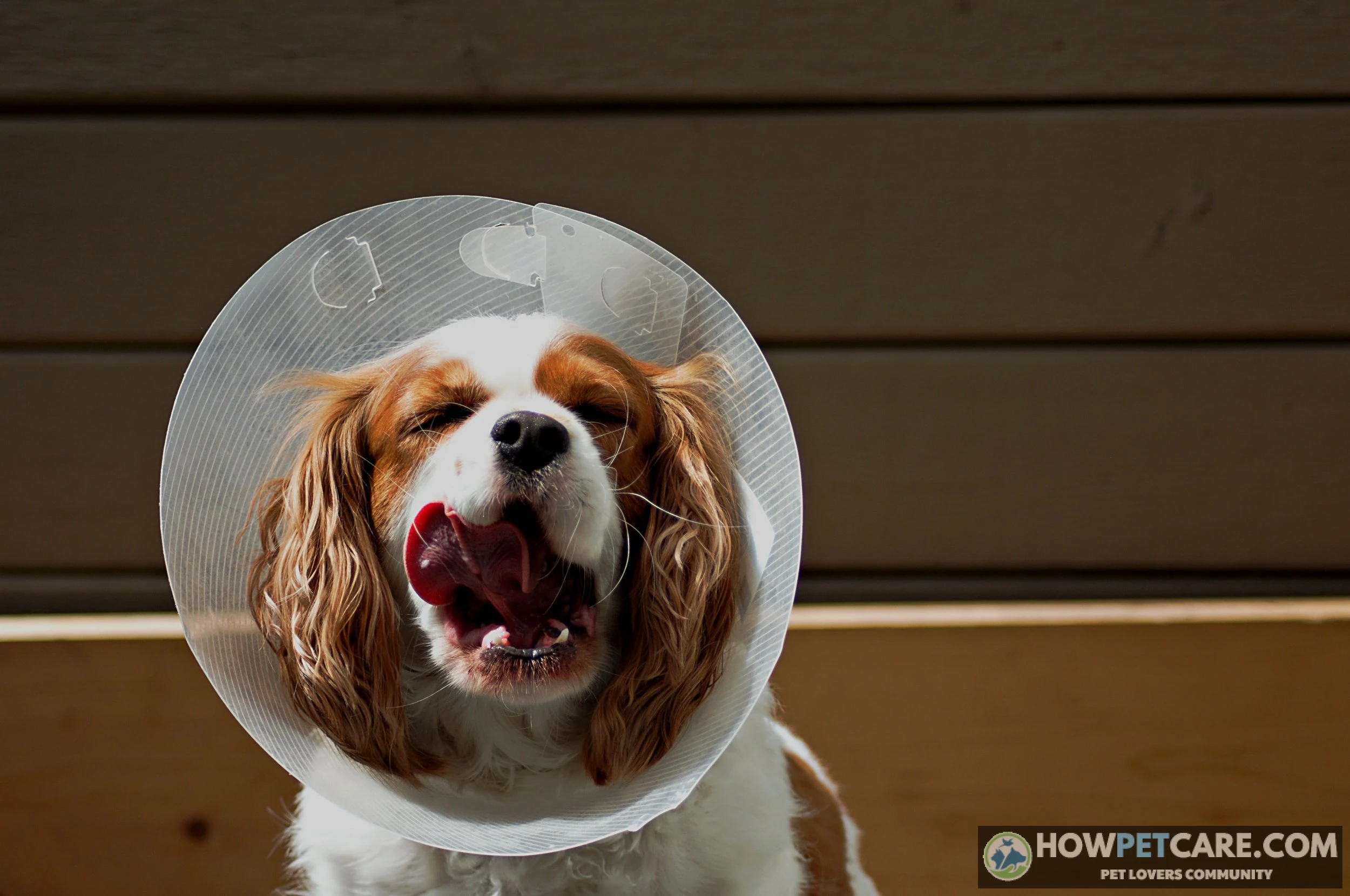

Hot spots on dogs are rapidly developing, wet, inflamed skin lesions caused by intense licking, chewing, or scratching. Immediately prevent contact with an Elizabethan collar, gently clean the area with sterile saline or properly diluted chlorhexidine, keep it uncovered and dry, and seek veterinary care for spreading, painful, deep, foul-smelling, or facial lesions.
A hot spot on a dog—clinically classified as acute moist dermatitis or pyotraumatic dermatitis—is a sudden, intensely itchy, inflamed, and weeping bacterial skin infection caused by rapid self-trauma. If you have just spotted a raw, red patch that appeared almost out of nowhere, focus on physical stabilization first: stop the scratching immediately, clean the surface with a non-stinging antiseptic, and determine whether the wound requires a same-day veterinary visit.
Hot spots on dogs can double in surface area in mere hours. Prompt intervention keeps a superficial irritation from turning into a deep, painful structural infection.
First-24-Hours Triage: Immediate First Aid for Canine Hot Spots
When a dog develops an acute hot spot, frantic licking, chewing, and scratching create an aggressive itch-scratch cycle. Surface bacteria, predominantly normal skin microflora like Staphylococcus pseudintermedius, rapidly colonize the micro-tears in the epidermal barrier. Stopping this self-trauma is the single most critical step in first aid.
Halting the Self-Trauma Cycle: Cones and Barriers
Before gathering supplies or inspecting the skin, put a physical barrier on your dog. A standard plastic Elizabethan collar remains the most reliable option. Inflatable donut collars seem comfortable, but flexible dogs can readily bend around them to chew their flanks, paws, or tail base. If the lesion sits on the torso or belly, a surgical recovery suit or a snug cotton T-shirt secured with medical tape works well while you locate a cone.
Never bandage or wrap a hot spot. Covering acute moist dermatitis with adhesive wraps or non-breathable gauze traps moisture, elevates local skin temperature, and creates an ideal anaerobic environment for bacterial proliferation. Exposure to dry, circulating air is essential for resolution.

Never use human household scissors to trim matted fur around an active hot spot. Inflamed canine skin swells unevenly and adheres tightly to damp fur. A flinching, painful dog can easily shift, leading to severe full-thickness skin lacerations that require surgical suturing. Use only professional electric clippers with a guarded blade (such as a #10 blade), or leave clipping entirely to veterinary staff.
Safe Cleaning Protocol: Antiseptic Dilutions and Technique
Once the barrier is secure, focus on gentle cleansing to remove crusted exudate and lower the microbial load without causing tissue necrosis.
-
Secure physical restraint and barrier
Fit an Elizabethan collar to prevent the dog from biting or flinching during treatment. Have a second person gently hold and calm the dog.
-
Carefully expose the lesion
If you own electric pet clippers with a safe guard, trim fur 1 inch around the margins to allow airflow. If specialized clippers are unavailable, do not use scissors; leave the fur intact and proceed directly to washing.
-
Prepare safe antiseptic
Use sterile saline solution (0.9% NaCl) or dilute chlorhexidine gluconate to a 2% concentration (diluting standard 4% solution 1:1 with distilled water). Avoid alcohol, witch hazel, or hydrogen peroxide, which destroy healing tissue.
-
Pat the surface gently
Saturate a sterile gauze pad with the solution. Dab the weeping skin gently to lift away crusts. Never rub or scrub the raw dermis.
-
Apply a soothing compress
Place a clean, cool, damp washcloth against the lesion for 5 to 10 minutes to alleviate heat and acute pain. Pat completely dry with a lint-free towel.

Identifying Dog Skin Hot Spots: Clinical Appearance vs. Lookalike Conditions
Acute moist dermatitis presents with a distinct clinical picture, but several other canine skin conditions present with hair loss, scabs, or redness. Misidentifying the issue risks the wrong treatment—such as applying antibacterial washes to fungal ringworm or mistaking parasitic mange for simple allergic scratching.
True hot spots develop fast. Skin can transform from normal tissue into a bright red, oozing lesion within 2 to 6 hours. Lesions are tender to the touch, fiercely itchy, and feature wet fur glued flat by discharge. Common sites include the neck, cheeks, base of the tail, flanks, and lateral thighs.

| Condition | Appearance | Moisture Level | Hair Loss Pattern | Onset Speed | Contagious Risk |
|---|---|---|---|---|---|
| Hot Spot (Pyotraumatic Dermatitis) | Bright red, raw, eroded skin; often weeping yellow or clear serum | Wet, actively oozing exudate | Focal patch, surrounding fur matted by discharge | Rapid (hours) | Non-contagious (underlying cause like fleas may spread) |
| Ringworm (Dermatophytosis) | Circular, scaly, erythematous border; central clearing | Usually dry; minimal exudate | Well-demarcated circular alopecia with broken stubble | Slow (days to weeks) | Highly contagious to pets and humans (zoonotic) |
| Sarcoptic / Demodectic Mange | Papules, yellowish crusts, lichenified (thickened) skin | Dry to greasy; rarely weeping unless secondary pyoderma | Generalized or patchy; common on ear margins and hocks | Progressive over weeks | Sarcoptic is highly contagious; Demodectic is non-contagious |
| Acral Lick Granuloma | Hard, raised, ulcerated fibrous nodule or plaque | Firm, chronic serous surface; not acutely wet | Localized plaque, hair stripped by persistent licking | Indolent (months of chronic licking) | Non-contagious |
Acute Moist Dermatitis vs. Fungal Ringworm
While an acute hot spot represents an explosive bacterial flare-up driven by self-trauma, ringworm (dermatophytosis) is a localized fungal infection of the hair follicles and outer skin layer. Ringworm develops slowly over several days to weeks. The classic presentation features expanding circular rings of brittle hair with fine scaling or gray crusts. Itching in ringworm ranges from absent to mild, whereas a dog with a hot spot displays frantic pruritus. Ringworm is zoonotic, meaning it readily transfers to humans and other pets. Hot spots cannot spread directly to other animals or people.
Mange and Chronic Lick Granulomas: Spotting the Differences
Parasitic mange, particularly sarcoptic mange (scabies), generates intense pruritus, but it manifests differently than an isolated hot spot. Mange presents with diffuse red bumps, dry yellowish crusting, and progressive hair loss concentrated along the ear pinnae margins, hocks, and elbows. Sarcoptic mange mites are contagious and will gradually colonize other animals in the household.
Acral lick granulomas, by contrast, are chronic, thickened plaques commonly found on the lower limbs (carpus or metacarpus). Unlike the sudden, weeping fire of an acute hot spot, a lick granuloma represents months of repetitive licking driven by orthopedic pain, deep-seated furunculosis, or behavioral anxiety, resulting in scar tissue and dense fibrosis.

Triage Checklist: Safe Interim Home Care vs. Same-Day Vet Emergency
Not every moist dermatitis lesion requires an emergency clinic visit. However, superficial hot spots can progress into deep pyotraumatic folliculitis or furunculosis, where bacterial infection ruptures hair follicles and invades subcutaneous tissue. Use this rubric to determine whether your dog can wait until morning or needs immediate clinical care.
Red-Flag Symptoms Requiring Emergency Care
- Facial Proximity: The lesion sits within 2 inches of the eye, directly around the mouth, or tracks into the ear canal. Facial swelling can compromise ocular structures or signal severe otitis externa requiring deep canal lavage.
- Signs of Systemic Infection: Rectal temperature exceeds 103.5°F (normal canine range is 100.5°F–102.5°F), accompanied by lethargy, anorexia, tremors, or refusal to drink.
- Structural Tissue Breakdown: Skin appears deeply ulcerated, displays crater-like pockets, oozes thick bloody or grayish-green purulent exudate, or emits a rancid, necrotic odor.
- Rapid Uncontrolled Expansion: Lesion spans greater than 3 to 4 inches in diameter or expands visibly over an 8-hour period despite barrier enforcement.
- Extreme Guarding or Pain: The dog growls, snaps, trembles, or enters severe distress when anyone approaches the affected area, indicating the need for professional chemical analgesia.
Criteria for Safe Overnight Monitoring
- The lesion is small (under 2 inches in diameter), superficial, and bright red with only clear to pale-yellow serous discharge.
- The dog remains alert, happily accepts food and water, and shows no systemic depression.
- An Elizabethan collar is securely in place, completely preventing the dog from making contact with the site.
- The lesion does not smell foul or show deep subcutaneous tracking.
Home stabilization is strictly an interim measure to protect the tissue while you arrange professional veterinary care. Any hot spot that fails to show distinct signs of drying and improved margin stability within 24 hours demands an in-person veterinary exam.

The Dangers of Kitchen-Cabinet Hacks and Human Over-the-Counter Medications
When faced with an angry, red skin patch, many well-meaning pet owners turn to viral home remedies or human medicine cabinets. Canine skin differs substantially from human skin: it is thinner (3–5 cell layers versus 10–15 cell layers in humans), maintains a neutral pH (around 6.8 to 7.4, compared to human skin at 5.5), and reacts aggressively to contact irritation.
Why Vinegar, Tea Tree Oil, and Coconut Oil Backfire
- Apple Cider Vinegar (ACV): Undiluted or even mildly diluted ACV is an acid (pH 2–3). Pouring acid onto raw, ulcerated skin stripped of its protective stratum corneum causes intense burning, severe pain, and local tissue damage that deepens the wound.
- Tea Tree Oil (Melaleuca Oil): Tea tree oil contains high concentrations of terpinen-4-ol and 1,8-cineole. Dogs absorb these compounds rapidly through damaged skin. Topically applied tea tree oil can induce neurotoxicity characterized by ataxia (loss of coordination), muscle tremors, hypothermia, depression, and elevated liver enzymes. It should never be applied to canine skin.
- Coconut Oil: While touted for mild antimicrobial properties in laboratory settings, applying coconut oil to a weeping hot spot creates an occlusive, oxygen-deprived seal. This greasy barrier locks in heat and moisture, accelerates skin maceration, and creates an optimal environment for rapid bacterial multiplication.
Neosporin, Hydrocortisone, and Benadryl: Toxicity and Efficacy Limits
- Triple Antibiotic Ointment (Neosporin): Standard formulations contain bacitracin, neomycin, and polymyxin B in a petroleum base. If your dog licks this ointment, neomycin can trigger severe gastrointestinal irritation and vomiting. Petroleum products also trap wound exudate. Never apply zinc-based diaper creams (like Desitin); ingested zinc induces life-threatening intravascular hemolytic anemia in dogs.
- Over-the-Counter Hydrocortisone 1%: While occasionally applied 3 to 4 times daily under strict veterinary direction, unsupervised topical steroid use carries major risks. Topical steroids inhibit local immune defense. If applied to a deep bacterial or fungal infection, hydrocortisone allows pathogens to multiply unchecked. Systemic absorption through licking can suppress adrenal function as well.
- Oral Diphenhydramine (Benadryl): Diphenhydramine is an H1-receptor antihistamine. While widely used for acute histamine-driven allergic reactions (like bee stings or vaccine reactions), it is notoriously ineffective at stopping the intense pruritus driven by established bacterial pyoderma, interleukin-31 pathways, or deep folliculitis. Relying on Benadryl delays proper antimicrobial care and leaves the dog in active distress.
Never give your dog human over-the-counter pain medications such as ibuprofen (Advil), naproxen (Aleve), or acetaminophen (Tylenol). These compounds induce severe gastric ulceration, acute renal failure, and fatal hepatic necrosis in dogs. Only a licensed veterinarian can prescribe safe, canine-specific non-steroidal anti-inflammatory drugs (NSAIDs) like carprofen or meloxicam.

What to Expect at the Veterinary Clinic: Procedures, Prescriptions, and Realistic Costs
A veterinary visit transforms an agonizing condition into an organized, rapid-healing process. Knowing what happens during the appointment clarifies the value of diagnostic steps and prepares you for the costs.
Diagnostic Cytology and Prescription Therapeutics
Upon intake, the veterinary team conducts an initial physical exam. If the lesion is intensely painful, the veterinarian may recommend light sedation or local topical lidocaine sprays so the area can be clipped and cleaned humanely.
Medical-grade electric clippers shave a wide perimeter around the lesion, usually 1 to 2 inches beyond all visible edges. Removing matted fur immediately exposes the skin to air, halting the moist maceration process.
Next, the clinician performs an impression cytology: pressing a clean glass microscope slide or transparent adhesive tape directly against the weeping dermis. Staining and examining this slide under the microscope reveals the exact microbial population: whether the infection is dominated by cocci bacteria (Staphylococcus), rod-shaped bacteria (Pseudomonas), or opportunistic Malassezia yeast organisms. Cytology ensures targeted medications rather than guesswork.
Prescription therapy typically follows a multi-modal approach:

- Targeted Topicals: Veterinary prescription sprays or ointments combining a topical antibiotic with a potent anti-inflammatory (such as gentamicin with betamethasone dipropionate) applied twice daily to halt local microbial growth and soothe inflammation.
- Targeted Antipruritics: Rapid-acting anti-itch interventions, such as a low-dose tapering course of oral prednisone, an injection of lokivetmab (Cytopoint), or oclacitinib (Apoquel), which turn off the neurological itch signaling cascade within hours.
- Systemic Antibiotics: Reserved for cases of pyotraumatic folliculitis or furunculosis where infection extends into deep tissue layers. First-line oral choices include cephalexin, cefpodoxime, or amoxicillin-clavulanate, usually prescribed for 14 to 21 consecutive days.
Itemized Treatment Cost Breakdown
Veterinary medical costs vary by region, clinic type (general practice versus 24-hour emergency hospital), and pet size. Below is a realistic out-of-pocket pricing breakdown reflecting typical veterinary fee schedules in 2026.
| Service / Medication | Low Estimate | High Estimate | Clinical Purpose |
|---|---|---|---|
| Physical Examination | $60 | $120 | Full systemic physical assessment and triage |
| Skin Surface Cytology & Microscopic Staining | $35 | $65 | Identifies specific bacteria (cocci/rods) or yeast overgrowth |
| Medical Fur Clipping & Antiseptic Lavage | $25 | $60 | Clears debris, exposes margins, promotes air drying |
| Mild In-Clinic Sedation / Analgesia (if required) | $75 | $180 | Provides humane pain relief for severe or sensitive lesions |
| Prescription Topical Medication (Spray/Cream) | $30 | $70 | Delivers localized antimicrobial and anti-inflammatory therapy |
| Systemic Oral Antibiotics (e.g., Cephalexin) | $40 | $110 | Clears deep tissue follicular infection |
| Targeted Antipruritic (Prednisone, Apoquel, or Cytopoint) | $35 | $95 | Shuts down itch reflex within 4 to 12 hours |
| Medical Elizabethan Collar | $15 | $45 | Prevents self-mutilation during the healing phase |
| Total Estimated Investment | $315 | $745 | Comprehensive diagnostic and clinical resolution |
Root-Cause Eradication and Long-Term Prevention Strategy
An acute hot spot is rarely an isolated problem; it is a physical reaction to an underlying trigger that caused relentless scratching in the first place. Treating the current lesion without addressing that initial trigger virtually guarantees recurrence.
Targeting the Underlying Triggers: Fleas, Ears, and Allergies
- Flea Allergy Dermatitis (FAD): The bite of a single flea injects antigenic saliva that triggers severe, widespread pruritus in allergic dogs. Hot spots clustered over the lower back, lumbar spine, and tail base are classic signs of flea allergy. Maintaining continuous, year-round administration of modern prescription oral flea prevention (such as the isoxazoline class: fluralaner, sarolaner, or afoxolaner) is the single most effective preventive measure.
- Otitis Externa (Ear Infections): Hot spots located beneath the ear, along the jawline, or on the lateral neck are almost invariably sparked by an underlying ear infection. Pain and inflammation deep in the horizontal ear canal lead the dog to violently scratch the side of the neck with its hind paws, lacerating the skin and creating a hot spot within hours. Whenever a neck hot spot appears, a thorough otoscopic exam is required.
- Undercoat Moisture and Matting: Dense-coated, double-coated, and water-retrieving breeds—notably Golden Retrievers, German Shepherds, Labradors, and Newfoundlands—face an elevated risk of acute moist dermatitis. When these dogs swim or get bathed, their dense undercoats can trap water against the warm skin for days. This prolonged dampness causes epidermal maceration, weakening cellular junctions and allowing surface bacteria to invade. High-velocity forced-air blow drying is essential after swimming or bathing for double-coated breeds.
- Anal Gland Impaction: Intense chewing around the tail base, perineum, or posterior thighs often stems from full or infected anal sacs rather than a primary skin issue. Routine veterinary checks help identify and express full glands before chronic licking sparks open sores; checking the perianal region at least once weekly is a recommended habit.
Coat Maintenance and Barrier Support for High-Risk Breeds
To build resilient skin that resists secondary bacterial invasion, institute a proactive skin-barrier health protocol:

- High-Dose Omega-3 Fatty Acids: Supplement your dog’s diet with veterinary-formulated marine lipid oils providing adequate therapeutic doses of eicosapentaenoic acid (EPA) and docosahexaenoic acid (DHA). Omega-3s incorporate into cellular membranes, downregulating systemic inflammatory cascades and reinforcing the lipid matrix of the stratum corneum.
- Ceramide and Phytosphingosine Washes: Bathe allergy-prone dogs with shampoos and microemulsion sprays containing synthetic ceramides and antimicrobial ingredients like 2% chlorhexidine. Ceramides replenish the intercellular lipids that prevent micro-cracking and bacterial penetration.
- Rigorous Undercoat Raking: Regularly use undercoat de-shedding rakes to remove dead, shed hair trapped beneath the topcoat. Removing this dense, dead wool permits air to circulate freely across the skin surface, preventing localized moisture pockets from forming.
Healing Milestones and Monitoring Progression
Hot spots themselves are strictly non-contagious. The lesion is simply an overgrowth of your dog’s own natural skin bacteria driven by mechanical self-trauma; it cannot transfer to other dogs, cats, or humans. If the primary trigger happens to be an ectoparasite—such as fleas or sarcoptic mange mites—that parasite can spread.
Managed properly with a barrier collar and appropriate veterinary therapeutics, pyotraumatic dermatitis follows a predictable healing timeline:
- Days 1 to 2 (Drying Phase): Under the influence of airflow and topical or systemic medications, the active oozing halts. The glossy, wet appearance transitions into a dry, flat surface. Margin expansion stops completely.
- Days 3 to 5 (Crusting Phase): The lesion forms a firm, dry, dark scab across the surface. Swelling and surrounding skin redness diminish significantly. The dog shows noticeably less urge to scratch when the collar is adjusted.
- Days 6 to 9 (Epithelialization Phase): Healthy new pink skin begins forming beneath the scab. The margins of the scab begin lifting and flaking away naturally. Never peel or pick these scabs off manually, as premature removal re-injures delicate new skin layers.
- Days 10 to 14 (Resolution and Hair Regrowth): The crusts slough off entirely, leaving smooth, slightly pink or hyperpigmented skin. Fine, soft hair stubble emerges across the formerly bald patch. Full coat density typically restores over the following 3 to 6 weeks.
By securing a cone immediately, avoiding caustic kitchen remedies, and partnering with your veterinarian to treat both the infection and its underlying spark, you can rapidly guide your dog from acute distress back to comfortable, healthy skin.

| Condition | Clinical Appearance | Moisture Level | Hair Loss Pattern | Onset Speed | Contagious Risk | Commonly Affected Areas | Underlying Cause or Pathogen |
|---|---|---|---|---|---|---|---|
| Hot Spot (Pyotraumatic Dermatitis) | Bright red, raw, eroded skin; often weeping yellow or clear serum | Wet, actively oozing exudate | Focal patch, surrounding fur matted by discharge | Rapid (hours) | Non-contagious (underlying cause like fleas may spread) | Neck, cheeks, base of the tail, flanks, and lateral thighs | Rapid self-trauma; surface bacteria (Staphylococcus pseudintermedius); underlying triggers (fleas, ear infections, moisture/matting, anal gland impaction) |
| Ringworm (Dermatophytosis) | Circular, scaly, erythematous border; central clearing; fine scaling or gray crusts | Usually dry; minimal exudate | Well-demarcated circular alopecia with broken stubble | Slow (days to weeks) | Highly contagious to pets and humans (zoonotic) | Not in source | Localized fungal infection of the hair follicles and outer skin layer |
| Sarcoptic / Demodectic Mange | Papules, yellowish crusts, lichenified (thickened) skin; diffuse red bumps | Dry to greasy; rarely weeping unless secondary pyoderma | Generalized or patchy; common on ear margins and hocks | Progressive over weeks | Sarcoptic is highly contagious; Demodectic is non-contagious | Ear pinnae margins, hocks, and elbows | Parasitic mange mites (Sarcoptes / Demodex) |
| Acral Lick Granuloma | Hard, raised, ulcerated fibrous nodule or plaque | Firm, chronic serous surface; not acutely wet | Localized plaque, hair stripped by persistent licking | Indolent (months of chronic licking) | Non-contagious | Lower limbs (carpus or metacarpus) | Months of repetitive licking driven by orthopedic pain, deep-seated furunculosis, or behavioral anxiety |
FAQ
Are hot spots on dogs contagious to other pets or humans?
No, acute hot spots are not contagious. They are secondary bacterial overgrowths, typically involving normal flora such as Staphylococcus pseudintermedius, caused by self-trauma; however, an underlying parasite such as fleas or sarcoptic mange can spread to other animals.
How long does a dog hot spot take to heal?
With proper treatment and an Elizabethan collar preventing self-trauma, a superficial hot spot typically dries within 48 to 72 hours and fully re-epithelializes within 7 to 10 days. Deep folliculitis or secondary infections may take 2 to 3 weeks to resolve completely.
Can a dog’s hot spot heal on its own without medication?
A very mild, early-stage surface hot spot can occasionally resolve if all licking is stopped with a cone and the skin is kept clean and dry. Most lesions expand rapidly, however, and may require veterinary-prescribed topical or oral antimicrobial treatment.
Why does my dog keep getting hot spots in the exact same spot?
Recurrent hot spots in one area suggest an unresolved underlying problem beneath or near the skin. Possible causes include a chronic ear infection, impacted anal glands, localized arthritis, allergies, or dense matting that repeatedly traps moisture.