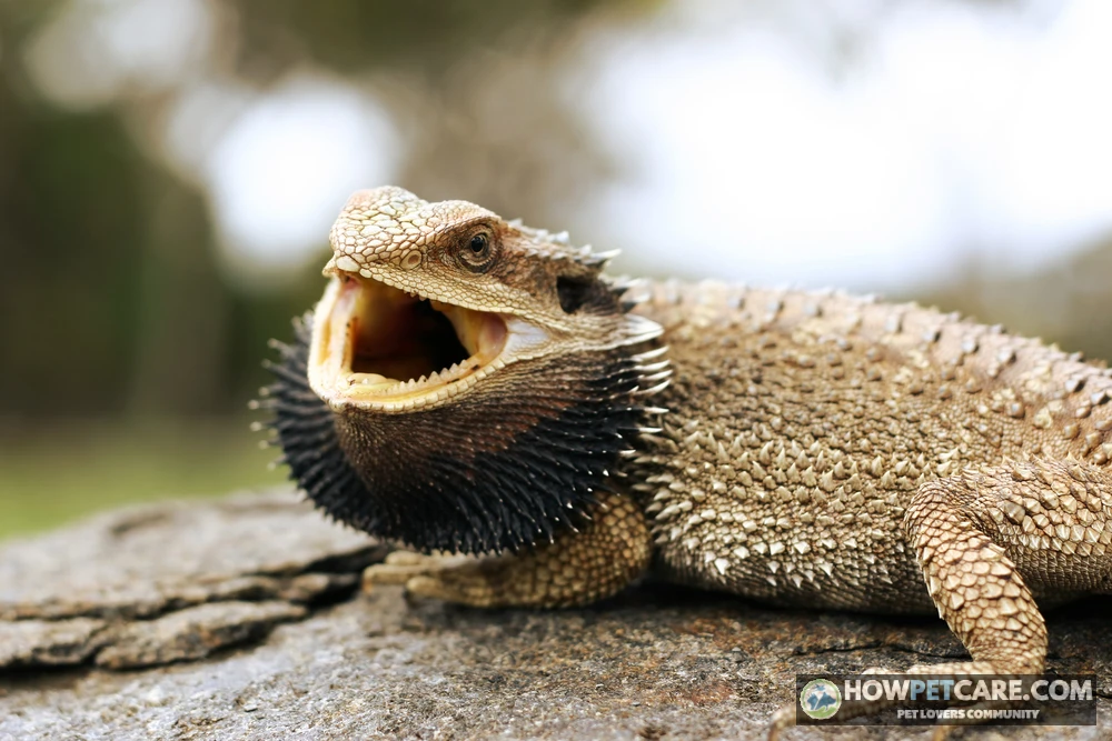

Bearded dragon mouth rot (infectious stomatitis) is a bacterial or fungal infection of the gums, lips, and jaw, usually triggered by poor temperatures, weak UVB, or injury. It never heals on its own — yellow plaques, swelling, or appetite loss require an exotics vet within 24–72 hours.
Bearded dragon mouth rot, clinically called infectious stomatitis, is a progressive bacterial or fungal infection that affects the gums, lips, oral mucous membranes, and jawbones. It occurs when an underlying problem—most commonly improper enclosure temperatures, failing UVB lighting, unhygienic conditions, or physical trauma—weakens the reptile’s immune system and allows opportunistic oral bacteria to invade. Bearded dragons have acrodont dentition: their teeth attach directly to the crest of the jawbone, with very little gum tissue in between. As a result, mild surface inflammation can quickly reach the underlying bone. Untreated mouth rot leads to deep tissue necrosis, permanent jaw deformities, systemic sepsis, and death. It won’t resolve on its own. A qualified reptile veterinarian needs to evaluate and treat it promptly.
This educational guide has been reviewed against current clinical reptile veterinary references. It can help owners identify symptoms, gauge urgency, and understand professional treatment protocols, but it isn’t a formal veterinary diagnosis or individualized medical instruction. If your bearded dragon shows signs of oral infection, pain, or systemic illness, consult an Association of Reptile and Amphibian Veterinarians (ARAV) licensed practitioner immediately.
Bearded Dragon Mouth Rot (Infectious Stomatitis): What It Looks Like and the Signs to Watch For
Infectious stomatitis isn’t an airborne infection that strikes out of nowhere; it develops secondary to immunosuppression or oral injury. A healthy bearded dragon’s mouth is lined with smooth, pale pink or flesh-colored tissue. When abrasions or chronic husbandry problems weaken the mucosal barriers, common opportunistic gram-negative oral bacteria, such as Aeromonas hydrophila and Pseudomonas aeruginosa, multiply uncontrollably and attack the gum line.
Early recognition matters. Initial lesions can be subtle and hidden behind the lip margins, while advanced disease involves extensive tissue destruction.
Early signs of mouth rot in bearded dragons
Infectious stomatitis can go unnoticed at first unless you inspect your bearded dragon’s mouth during regular handling. Early signs include:
- Pinpoint gingival erythema: Tiny red spots, subtle petechiae, or a thin red line where the teeth meet the gums.
- Mild localized swelling: Slight puffiness along one side of the lip or jaw, which may make the closed mouth look uneven.
- Stringy, viscous saliva: Normal saliva is clear and thin. Early infection can cause slightly tacky, cloudy, or stringy mucus when the dragon opens its mouth.
- Subtle behavioral hesitation: A dragon may approach food eagerly but chew awkwardly, favor one side of its mouth, drop hard-bodied insects, or pause between bites because its mouth is tender.
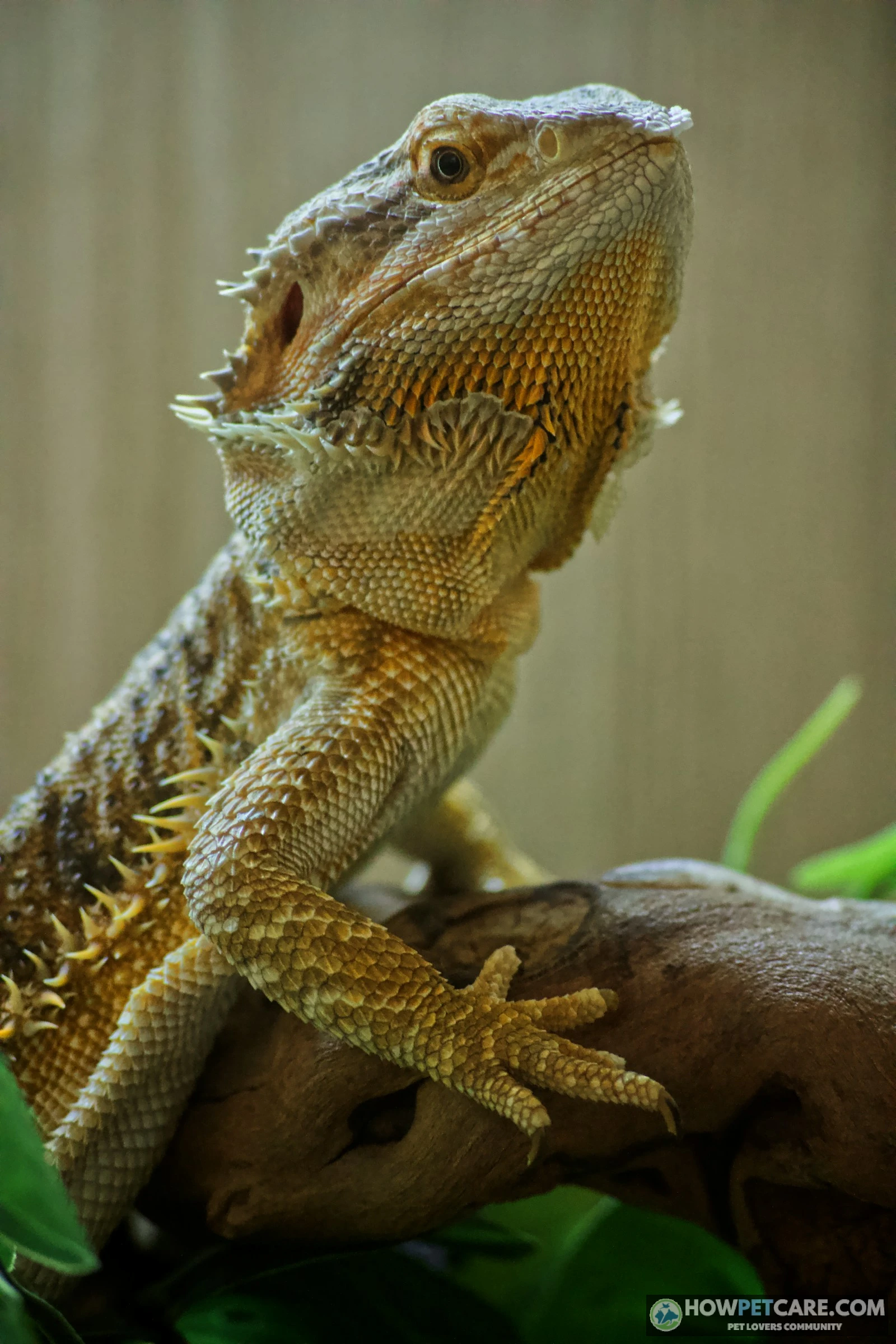
At this stage, your dragon may still have normal energy and body weight, making it easy to mistake the problem for something harmless.
Advanced mouth rot: what stage three and beyond looks like
As opportunistic bacteria move deeper into the oral mucosa and spaces beneath the gums, the immune response creates thick, necrotic debris. Reptilian heterophils—the reptile counterpart to mammalian neutrophils—lack the specific enzymes needed to liquefy dead cells. Reptilian pus therefore doesn’t drain as a liquid; it forms solid, caseous material resembling dry cottage cheese or curdled milk.
In advanced stages (stage three and beyond), signs include:
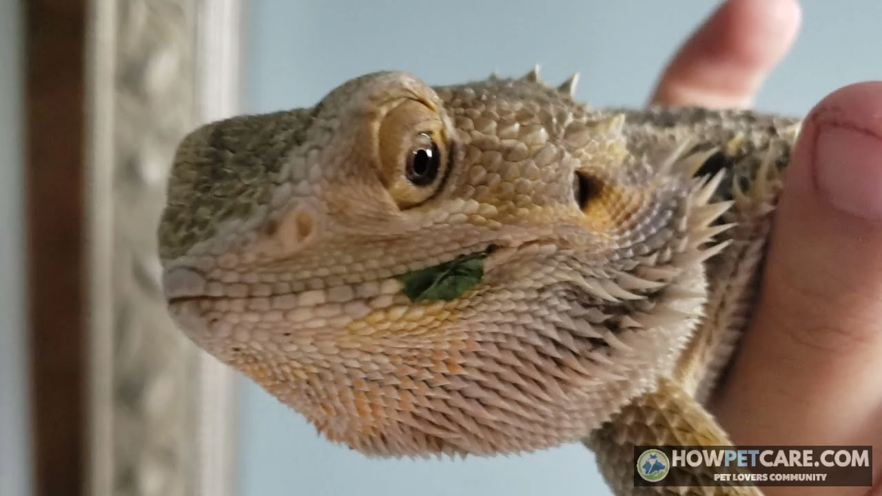
- Yellow or grayish-white cheesy plaques: Solid clumps of caseous exudate cling firmly to the gums, inner lips, palate, and tongue base. Scraping them off exposes raw, bleeding ulcerations underneath.
- Copious, thick oral discharge: Frothy, gray, or yellowish fluid bubbles from the corners of the lips or forms heavy strings across the mouth.
- Severe swelling and jaw deformity: The lower or upper jaw looks thickened, bulbous, or crooked as bacteria erode through the thin gingiva into the mandibular or maxillary bone tissue.
- Loose or shed teeth: Because acrodont teeth are fused to bone, osteomyelitis (bone infection) causes teeth to loosen, blacken, or fall out entirely.
- Severe halitosis: A distinct, foul, putrid odor comes from the mouth.
- Anorexia and lethargy: Severe pain leads to complete food refusal, accompanied by dark stress coloration (a persistently black beard), sunken eyes, and extreme weakness.
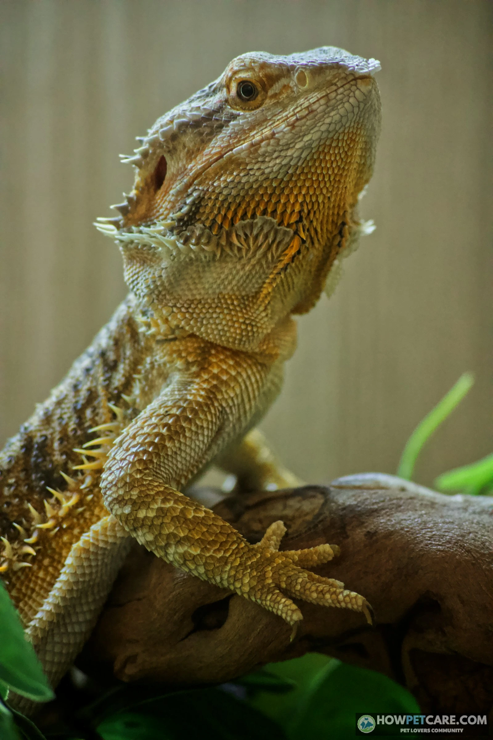
What a healthy bearded dragon mouth looks like
Knowing the normal oral anatomy of Pogona vitticeps can help you spot early changes:
- Color: The oral mucosa, inner gums, and palate are uniformly pale pink to light bubblegum pink. Some dragons naturally have flat, dusky gray-to-black melanin patches on the inner lip margins or tongue tip, but the underlying tissue is clean and uninflamed.
- Texture and moisture: Mucosal surfaces are glistening, moist, and smooth, without lumps, crusts, or flakes. Saliva is clear, thin, and watery, with no trailing strings or bubbling at the corners.
- Teeth and jaw alignment: Teeth look clean, creamy-white to light ivory, and firmly set in a continuous row along the jaw crest, without missing gaps, blackened roots, or tartar buildup. The upper and lower jaws close flush, without gaps, underbites, or lateral bulging.
Is It Mouth Rot or Something Else? Telling It Apart from Stuck Shed, Pigmentation, Burns, Trauma, and MBD
A discolored mark or swollen jaw isn’t always infectious stomatitis. The right response depends on the underlying problem, so distinguishing mouth rot from look-alikes helps prevent dangerous missteps.
Mouth rot versus normal pigmentation
Bearded dragons often have natural melanin pigmentation inside their mouths. These flat, smooth, symmetrical patches can appear as slate gray, blue-black, or deep purple spots along the inner gums, tongue, or roof of the mouth. Normal pigmentation doesn’t change in size or texture over weeks, produce discharge or swelling, or affect the dragon’s enthusiastic feeding behavior. Mouth rot lesions, by contrast, are raised, asymmetric, inflamed, and progress noticeably over time.
Mouth rot versus stuck shed
During ecdysis (shedding), dead skin around the snout, nostrils, and outer lip borders turns dull, papery, and whitish. Stuck shed is superficial and confined to the exterior scales. It doesn’t cause cheesy exudate, intraoral swelling, or an unpleasant odor. A gentle soak in lukewarm water softens stuck shed so it can lift naturally without bleeding. Mouth rot plaques are inside the mouth or directly on the mucosal gum tissue. They can’t be soaked away and bleed if disturbed.
Mouth rot versus trauma or burns
Physical injuries can resemble early stomatitis or provide a gateway for infection. Rostral abrasions occur when a stressed dragon repeatedly rubs its snout against glass enclosure walls or wire mesh, leaving a localized raw scrape or dry scab on the tip of the nose or outer lip. Thermal burns from overhead basking lamps, exposed ceramic heat emitters, or malfunctioning heat rocks produce sharply demarcated, blistered, or blackened lesions. Trauma and burns begin as sterile injuries, but open wounds inside the mouth can quickly be colonized by Pseudomonas or Aeromonas, turning the injury into active infectious stomatitis.
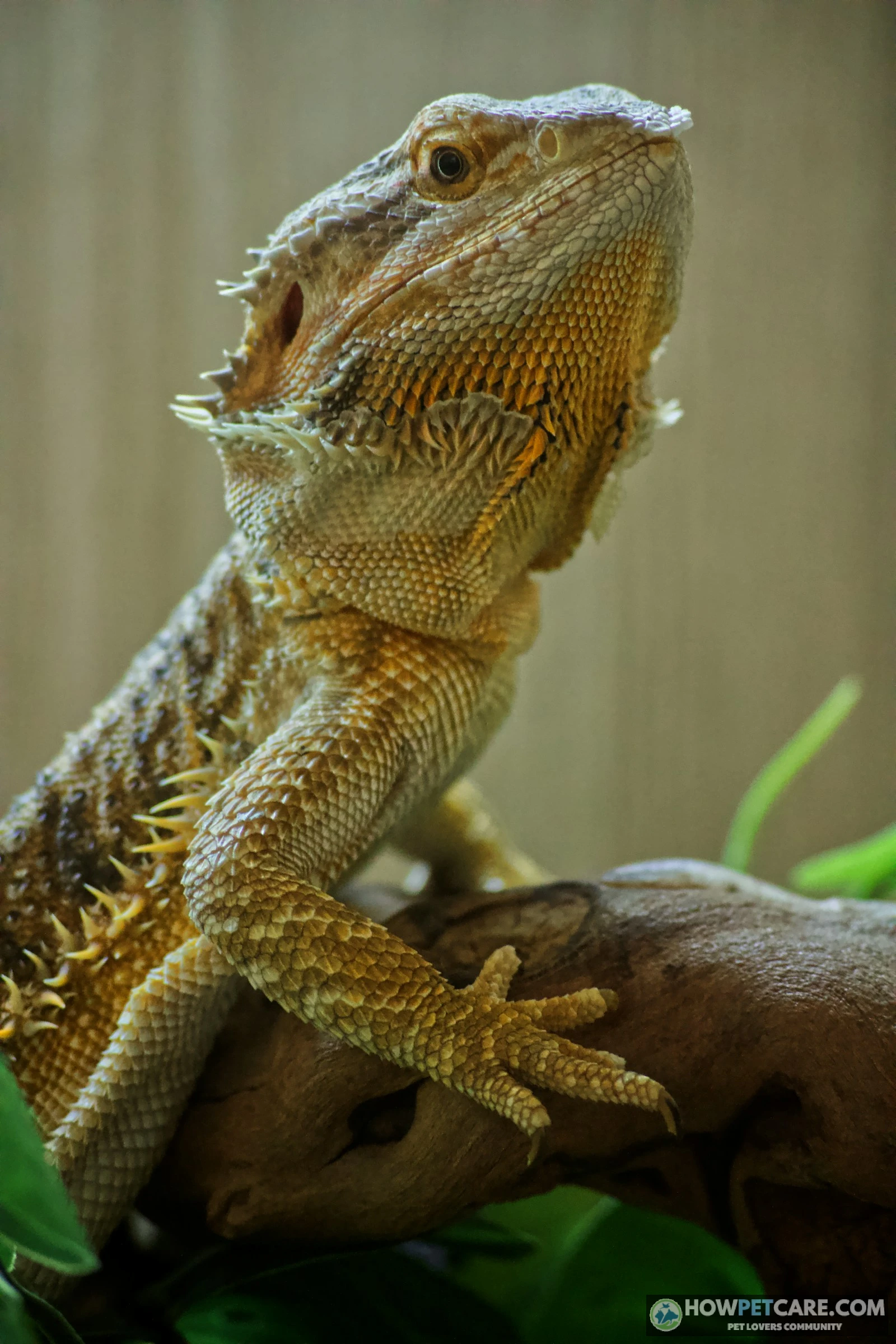
Swollen jaw: mouth rot or metabolic bone disease?
Metabolic Bone Disease (MBD) is caused by chronic calcium deficiency, vitamin D3 deficiency, or inadequate UVB lighting. In advanced MBD, the body leaches calcium from the skeleton, leaving the jawbones soft, flexible, and structurally thickened—a condition often called "rubber jaw." The lower jaw may look noticeably widened, foreshortened, or bowed outward.
Close inspection can help distinguish MBD from mouth rot:
- MBD jaw swelling is typically symmetrical, non-ulcerative, and firm or rubbery, without foul odor, intraoral discharge, or yellow plaques. Other signs often include twitching toes, tremors, bowed limbs, and difficulty lifting the body.
- Mouth rot swelling is usually asymmetric and soft to fluctuant, with intense mucosal redness, foul odor, and necrotic plaques.
MBD and mouth rot often occur together. Poor husbandry that causes metabolic bone disease also compromises the immune system, leaving the weakened jaw bone vulnerable to acute bacterial invasion. An exotics veterinarian needs radiographs (X-rays) to evaluate bone density and distinguish structural bone softening from osteomyelitis.
The table below compares the main differences:
| Condition | What it looks like | Where it appears | Discharge or plaques | Behavior and appetite | Other clues | Next step |
|---|---|---|---|---|---|---|
| Mouth rot (early) | Pinpoint red spots or reddened gum line, mild swelling, slightly stringy saliva | Inside the mouth: gums, lip margins, around teeth | None yet, or thin cloudy mucus | May chew oddly or hesitate at food; otherwise normal | Often follows an injury, cold spell, or dirty enclosure | Book a vet visit within the week |
| Mouth rot (advanced) | Yellow/white cheesy plaques, swollen or bleeding gums, loose teeth, foul smell | Gums, inner lips, jaw; may deform the jaw line | Thick pus or yellow plaques that don’t wipe away cleanly | Reduced appetite or refusal to eat, lethargy | Jaw may look swollen or misshapen | Same-day to 72-hour vet visit depending on eating status |
| Normal pigmentation | Flat dark grey, black, or brown patches | Gums, inner lips, roof of mouth | None | Completely normal | Symmetrical, unchanged for months/years, present since young | None — photograph and recheck monthly |
| Stuck shed | Dry, whitish, flaking or peeling skin | Outside the mouth: lips, snout, around the eyes | None inside the mouth | Normal | Softens and lifts after a warm soak; other shed patches on the body | Soak and monitor; vet check if a patch persists through two sheds |
| Rostral trauma (glass rubbing) | Raw scrape, scab, or worn scales | Tip of snout and upper lip, external | None, unless secondarily infected | Normal | Seen rubbing nose on glass; scrape is localized | Fix the stressor, keep clean, vet if it reddens or oozes |
| Thermal/chemical burn | Sharply defined raw or discolored patch | Area contacting a heat source or treated surface | None initially | May be normal or reduced | Linked to a new bulb, heat rock, or product exposure | Remove the source, vet visit — burns can become infected |
| MBD jaw swelling | Soft, rubbery, or bowed jaw; face may look swollen | Whole jaw structure, not just gums | Usually none — tissue isn’t oozing | May struggle to chew | Tremors, swollen limbs, bowed legs, poor UVB history | Vet visit with X-rays; MBD and mouth rot can occur together |
This table can help you decide how quickly to seek care, but it cannot replace a hands-on exam with specialized reptile mouth speculums. If you’re unsure between two rows, treat the situation as the more urgent condition.
 You might be wonderingMetabolic bone disease bearded dragonMetabolic Bone Disease in Bearded Dragons: Stages & Action PlanRead the answer
How Urgent Is It? Emergency, Same-Week Vet Visit, or Monitor at Home
Infectious stomatitis progresses across a spectrum. Whether your dragon needs emergency care today, an appointment this week, or short-term monitoring depends on its behavior and the extent of the lesions.
Use this triage matrix to match what you see to a next step:
| Urgency level | Signs you see | Why it matters | What to do now | Timeframe |
|---|---|---|---|---|
| Emergency | Open-mouth breathing, uncontrolled mouth bleeding, visibly deformed or broken jaw, total food refusal plus lethargy or collapse, or lesions spreading noticeably within 24 hours | Infection may have reached bone or become systemic; a crashed dragon can decline fast | Phone an exotics vet or emergency clinic now. Keep the dragon warm (normal basking temperature, not hotter) during transport | Same day — within hours |
| Same-week visit | Yellow or cheesy plaques, thick discharge, swollen gums, loose teeth, bad breath, reduced but not absent appetite | Active infection that will worsen but is stable enough for a scheduled appointment | Book an appointment, take dated photos tonight, fix temperature and UVB, isolate from other reptiles | Within 24–72 hours |
| Monitor — with a deadline | One small red spot or minor scrape; eating, activity, and droppings all normal | Could be early inflammation or a minor injury — but early mouth rot looks exactly like this | Photograph the spot, note the date, recheck daily | Vet visit if no improvement in 48 hours, or instantly if anything worsens |
Clinical Triage Ground Rules
Use these two rules when evaluating your dragon:
- "Monitoring" requires an explicit 48-hour expiration timer. Don’t wait passively for several weeks. If a tiny inflamed mark hasn’t resolved within 48 hours, or expands at all, schedule a veterinary consultation immediately.
- Appetite cessation automatically elevates your urgency by one full tier. A dragon with minor redness that suddenly stops eating is experiencing significant pain or systemic distress. Treat it as a same-week priority (within 24 to 48 hours), not a mild observation case.
What to Do Tonight While You Wait for the Vet Appointment
After scheduling your appointment, provide careful support during the 48-to-72-hour window before the visit. You can’t eradicate the infection at home. Your goal is to stabilize your pet, prevent secondary complications, and gather useful diagnostic records.
Temperature, UVB, and hydration checks
Reptiles are ectothermic, so their immune response, enzymatic pathways, and cellular defenses depend on environmental heat. Below its preferred optimum temperature zone, a bearded dragon’s heterophils and lymphocytes can’t mount an effective response against bacterial proliferation.
- Measure surface temperatures accurately: Use a calibrated infrared temperature gun or digital probe thermometer directly on the basking surface. For adult bearded dragons, check that the primary basking surface reaches 95°F to 105°F (35°C to 40.5°C). Keep the ambient cool side within 75°F to 85°F (24°C to 29°C). Maintain these ranges precisely: overheating causes thermal stress, while sub-optimal temperatures accelerate microbial invasion.
- Verify UVB output: Reptile immune systems require active vitamin D3 synthesis driven by adequate UVB exposure. Use a linear T5 HO (high output) fluorescent tube that covers one-half to two-thirds of the enclosure length. Confirm that the bulb is under 6 to 12 months old, mounted at the manufacturer’s recommended distance, and unobstructed by glass or plastic.
- Support hydration: Oral bacterial infections can cause dehydration through weeping lesions and reduced water consumption. Offer shallow, lukewarm water soaks (85°F to 90°F) up to the dragon’s shoulders for 10 to 15 minutes. Supervise closely and keep its head above water. You can also use a clean plastic pipette to offer clean water droplet by droplet on the tip of the snout, letting the dragon lick it voluntarily.
Isolating the dragon and keeping its enclosure clean
Secondary bacterial contamination flourishes in enclosures containing loose sand, bioactive soils, or textured bark. Particles can stick to exposed, ulcerated gums and introduce new pathogens directly into open tissue.
-
Transition to a hospital enclosure
Move the sick dragon into a clean, uncrowded enclosure or thoroughly strip its existing habitat. Remove all sand, wood chips, and particulate substrate. Line the entire floor with plain white paper towels or butcher paper. Replace the lining as soon as it’s soiled, and use it to monitor droppings and oral discharge.
-
Remove porous accessories
Remove natural driftwood, cork bark, and fabric hammocks. Porous decor can’t be fully sanitized and harbors opportunistic fungi and bacteria. Provide non-porous, easy-to-sanitize hides, such as smooth plastic reptile caves or clean ceramic tiles safely elevated on solid blocks.
-
Establish strict isolation
If you keep other reptiles, move the affected dragon into a separate room. Don’t share handling equipment, and wash your hands thoroughly after servicing the sick dragon’s enclosure.
Syringe feeding a dragon that refuses food: what to ask your vet first
A dragon with oral pain will often refuse crickets, roaches, and tough greens. Anorexia rapidly depletes liver glycogen reserves in reptiles. Force-feeding or improper syringe feeding, however, carries serious risks: oral trauma and fatal aspiration pneumonia if food enters the glottis, the airway opening visible on the floor of the dragon’s tongue.
Don’t attempt aggressive syringe feeding without veterinary approval. Before your appointment, call the clinic and ask:
- "My dragon has stopped eating; should I introduce an oral critical-care supplement (such as Oxbow Critical Care Herbivore or Fluker’s Repta+Boost) before my appointment?"
- "What specific daily volume (in milliliters) is safe for my dragon’s current body weight?"
If the veterinary team directs you to offer interim nutrition:
- Keep the dragon’s body naturally upright or angled slightly forward. Never tilt it onto its back.
- Gently express a tiny drop of pureed food onto the tip of the snout or lip margin and encourage the dragon to lick it voluntarily.
- If using a catheter syringe, introduce only a fraction of a milliliter along the lateral side of the mouth behind the front teeth, well away from the centrally located glottis.
- Stop immediately if the animal thrashes, gags, or shows signs of respiratory distress.
Giving the clinic organized details can help the team triage your appointment faster. Use this script when calling your exotics clinic:
"Hi, I have a bearded dragon, approximately [age], and I am concerned about an active oral infection. Inside the mouth, I observe [red spots / yellow caseous plaques / swelling / foul odor / loose teeth] located along the [left/right gums, inner lips, jaw crest]. I first identified this [number of days] ago, and it has [remained stable / expanded / worsened]. The dragon is currently [eating normally / hesitating on hard insects / completely refusing food for X days]. My basking surface temperature is [number]°F measured with an infrared temp gun, and my T5 HO UVB tube is [number of months] old. How soon can your exotics team evaluate him, and what interim stabilization steps do you recommend until then?"
Home remedies for bearded dragon mouth rot: what not to try
Online forums often suggest DIY treatments that can seriously damage delicate reptile tissue. Avoid these practices:
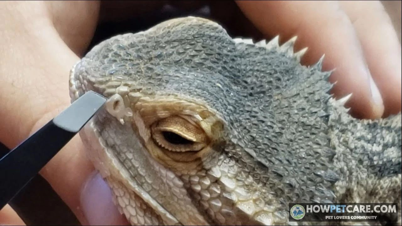
- Never pick, scrape, or debride oral plaques at home: Reptilian caseous exudate adheres tightly to fragile mucosal membranes and the underlying periosteum. Scraping it with cotton swabs, tweezers, or fingernails tears tissue, causes severe hemorrhaging, and forces opportunistic bacteria into the bloodstream, triggering fatal sepsis.
- Do not apply over-the-counter human ointments (Neosporin, Polysporin): Petroleum-based antibiotic ointments soften scales, destroy normal skin architecture, and become toxic when ingested. Many contain neomycin, which is nephrotoxic (damages kidneys) in dehydrated reptiles.
- Avoid human mouthwashes, hydrogen peroxide, and tea tree oil: Hydrogen peroxide destroys healthy epithelial cells along with bacteria, delaying wound healing. Human mouthwashes contain alcohol, flavorings, and xylitol, which are corrosive and toxic to reptiles. Pure tea tree oil and herbal extracts are rapidly absorbed through mucous membranes, leading to systemic neurotoxicity and organ failure.
- Do not guess chlorhexidine dilutions: Veterinary teams often prescribe chlorhexidine gluconate oral rinses, but the margin of safety is precise. Standard over-the-counter surgical scrub concentrations (typically 2% to 4%) chemically burn oral tissue. If your vet prescribes a clinical oral rinse, it requires precise dilution (typically down to 0.05%) and must be applied strictly as instructed.
What Causes Mouth Rot in Bearded Dragons, and the Husbandry Fix for Each Cause
Infectious stomatitis is rarely an unprovoked primary infection; it is almost universally a symptom of an underlying environmental deficiency or chronic physical stress. To prevent reinfection, check and correct each aspect of the dragon’s habitat.
Enclosure temperature and UVB lighting
- The Husbandry Failure: Chronic sub-optimal temperature is the most common trigger. When the basking zone or ambient enclosure falls below the preferred optimum temperature zone, the reptile’s immune system enters an energy-conservation mode. Heterophil phagocytosis drops, enzymatic tissue repair stalls, and naturally occurring oral flora become pathogenic. Weak or absent UVB radiation also halts cutaneous synthesis of cholecalciferol (vitamin D3), crippling calcium absorption and gut health.
- The Concrete Fix: Install a high-quality halogen basking bulb with a reliable dimming thermostat. Set the basking surface to maintain 95°F to 105°F (35°C to 40.5°C) for adult dragons, with a cool-side ambient zone of 75°F to 85°F (24°C to 29°C). Measure these zones daily with an infrared temperature gun. Discard outdated compact coil UVB bulbs and install an external fixture with a linear T5 HO 10.0 or 12% UVB tube spanning 50% to 70% of the enclosure. Replace the tube every 6 to 12 months, even if it still emits visible light; its phosphors degrade long before the bulb burns out.

Calcium deficiency, diet, and immunosuppression
- The Husbandry Failure: Chronic dietary calcium deficiencies or unbalanced phosphorus-to-calcium ratios lead to nutritional secondary hyperparathyroidism (MBD). As the skeleton softens, the alveolar ridges and mandibular margins lose mineral density, causing micro-fractures during chewing. Because bearded dragons’ acrodont teeth attach directly to the jawbone, this weakening allows oral bacteria to travel straight into osseous tissue.
- The Concrete Fix: For adults, feed a balanced rotational diet of 80% safe leafy greens and vegetables (collard greens, mustard greens, dandelion greens, turnip greens, butternut squash) and 20% healthy gut-loaded feeder insects (dubia roaches, black soldier fly larvae, crickets). Dust all feeder insects with a pure calcium carbonate supplement (without D3) at almost every feeding. Alternate with a high-quality multivitamin and calcium with D3 once or twice weekly, depending on UVB exposure levels.
Mouth injuries and scale rot as entry points
- The Husbandry Failure: Mucosal injuries give opportunistic pathogens a direct entry point. Live crickets left in an enclosure overnight will chew on the soft tissue of a sleeping dragon’s lips, eyelids, and vent. Dragons under environmental stress may also rub their snouts obsessively against clear glass (glass surfing), wearing down the rostral scales until bare, bleeding tissue is exposed.
- The Concrete Fix: Don’t leave uneaten live feeder insects in the habitat. Remove uneaten crickets or roaches within 15 minutes of feeding. To stop glass surfing, assess environmental stressors: cover the back and sides of glass terrariums with non-reflective backgrounds, verify enclosure dimensions (a minimum 4x2x2 foot / 120-gallon terrarium is standard for an adult), and provide appropriate thermal gradients and visual hides. Keep relative humidity strictly between 20% and 40%; excess dampness promotes fungal growth and scale rot, which can spread to oral margins.
How Vets Diagnose and Treat Bearded Dragon Mouth Rot: Antibiotics, Debridement, and Recovery
Knowing what to expect at an exotics veterinary consultation can ease anxiety and help you ask about diagnostics, medication courses, and recovery.
Step-by-step: what happens at the exotics vet visit
-
Comprehensive clinical evaluation
The veterinarian performs a physical exam, palpates the jawline, assesses body condition, and listens to the lung fields for secondary aspiration pneumonia. The vet also carefully opens the mouth with atraumatic soft speculums to examine the gingival folds, palate, and glottis.
-
Diagnostic cytology and bacterial culture
The practitioner gently collects a sample of oral exudate with a sterile swab. Immediate in-house cytology staining and microscopic examination identifies the broad pathogen class (gram-negative rods, cocci, or fungal hyphae). The swab then goes to an external veterinary laboratory for aerobic culture and antibiotic sensitivity testing.
-
Staging radiographs (X-rays)
For dragons with mandibular swelling, loose teeth, or deep lesions, orthogonal full-body radiographs are essential. X-rays show whether infection has penetrated the bone cortex, causing osteomyelitis, bone lysis, or pathological jaw fractures.
-
Surgical debridement
Under local sedation or light general anesthesia, the veterinarian gently debrides the mouth, removes caseous yellow plaques, flush-cleanses necrotic pockets with sterile saline, and extracts unsalvageable, mobile teeth that harbor deep bacterial colonies.
Which antibiotics are used for reptile mouth rot, and why culture matters
Infectious stomatitis in reptiles is primarily driven by gram-negative organisms such as Pseudomonas aeruginosa, Aeromonas hydrophila, Klebsiella, and Serratia. These bacteria have varied resistance patterns, making empiric antibiotic selection risky. A formal culture and sensitivity panel identifies the precise organism and lists the medications that will successfully eliminate it.
Depending on culture results and systemic health, a reptile veterinarian may prescribe medications from several drug classes:
- Cephalosporins: Injectable agents such as ceftazidime are widely used for their broad gram-negative coverage, good tissue penetration, and low organ toxicity.
- Fluoroquinolones: Drugs such as enrofloxacin provide potent bactericidal action and are given orally or by injection. They require careful hydration management to avoid renal stress.
- Aminoglycosides: For severe, resistant infections, medications such as amikacin may be used under close veterinary monitoring, typically with aggressive fluid therapy because of nephrotoxicity risks.
- Topical Antiseptics: These include professional flushes with precisely diluted chlorhexidine gluconate (0.05%) or silver sulfadiazine cream applied to debrided mucosal lesions.
- Analgesics and NSAIDs: Pain relief is vital. Systemic anti-inflammatories such as meloxicam reduce swelling and restore comfort, encouraging the dragon to resume voluntary feeding.
Never administer leftover antibiotics or obtain veterinary drugs without a prescription. Reptile dosages are heavily influenced by body weight, hydration, ambient temperature, and renal clearance rates. Improper administration causes kidney failure or fosters antibiotic resistance.
Can mouth rot heal on its own?
Some new reptile keepers believe mild mouth rot can clear up if they raise the temperature slightly. It can’t. Mouth rot does not heal without medical intervention.
Reptilian heterophils form solid, non-draining caseous plugs, so the body cannot purge the necrotic core on its own. Bacteria multiply behind this barrier and erode deeper into the periodontal ligament and jawbone. Without treatment by an exotics specialist, a small red mark will predictably progress into osteomyelitis, systemic bacteremia, and death.
Recovery timeline and recurrence risk by stage
Full recovery depends heavily on the stage at diagnosis and prompt correction of environmental deficiencies.
| Stage at diagnosis | Typical treatment window | What healing looks like | Husbandry fix required at this point |
|---|---|---|---|
| Early (gum redness, small lesions, no plaques) | ~4–8 weeks with medication and rechecks | Gums return to pink, appetite normalizes within 1–2 weeks of starting treatment | Find and fix the trigger now — temperature, UVB age, or hygiene — or the case relapses |
| Moderate (plaques, discharge, debridement needed) | 2–3+ months including follow-up visits | Plaques shrink and slough, discharge stops, eating resumes | Full enclosure deep-clean plus correction of the identified failure before the course ends |
| Advanced (bone involvement, jaw deformity, surgery) | 3–6 months, sometimes with permanent jaw changes | Guarded recovery; jaw may never look fully normal | Every husbandry factor must be corrected — recurrence with bone damage is harder to treat |
Signs of progress include oral tissues returning to pink, discharge stopping, plaque borders shrinking, and steady weight gain. Recurrence is common, though, if the underlying husbandry problem isn’t corrected. If basking temperatures or UVB levels return to substandard ranges after antibiotics end, opportunistic pathogens will reinfect the vulnerable gum line.
Bearded Dragon Mouth Rot Vet Cost: Itemized Exam, Testing, and Treatment Prices
The cost of specialized reptile care is a major concern for many owners. Exotics care requires specialized equipment and training, making procedural fees comparable to dog and cat medical services.
The ranges below reflect realistic 2026 United States veterinary clinic estimates for regional exotics practices and specialty hospitals:
| Service or item | What it tells or does | Typical cost range (USD) | Needed in early cases? | Needed in advanced cases? | Source and year |
|---|---|---|---|---|---|
| Initial exotics exam | Comprehensive physical assessment and oral evaluation | $80 – $200 | Yes | Yes | Exotics clinic fee survey (2026) |
| Oral cytology | Immediate microscopic assessment of cells and bacteria | $50 – $120 | Recommended | Yes | Exotics clinic fee survey (2026) |
| Bacterial culture & sensitivity | Identifies exact bacterial strain and effective antibiotics | $180 – $350 | Recommended | Yes | Veterinary reference lab standard pricing (2026) |
| Diagnostic X-rays (orthogonal) | Assesses jawbone density, osteomyelitis, and fractures | $150 – $320 | Rarely | Yes | Exotics radiology standard pricing (2026) |
| Sedation & surgical debridement | Light anesthesia, plaque removal, and oral flush | $200 – $500 | No | Yes | Specialty hospital surgical rates (2026) |
| Systemic antibiotics & pain meds | 4 to 8-week courses of injectables/orals (ceftazidime, meloxicam) | $60 – $180 | Yes | Yes | Exotics pharmacy pricing (2026) |
| Follow-up recheck exams | Visual reassessments and treatment monitoring | $60 – $120 (per visit) | Yes (1–2 visits) | Yes (3–5 visits) | Regional practice estimates (2026) |
Early-stage versus advanced-stage cost scenarios
Scenario A: Early-Stage Stomatitis ($400 – $900 total)
An owner notices localized gum redness and books an appointment within 48 hours. The visit includes an exotics consultation ($110), in-house cytology ($60), a culture and sensitivity panel ($220), a 4-week course of injectable ceftazidime and oral meloxicam ($95), and one follow-up recheck ($75). Total investment: approximately $560. Healing is rapid, with no bone damage or surgery needed.Scenario B: Advanced Chronic Stomatitis with Osteomyelitis ($1,100 – $2,500+ total)
The condition has progressed for weeks. Caseous yellow plaques cover the gums, the jaw is deformed, and the dragon has stopped eating. Care includes an emergency consultation ($195), comprehensive blood chemistry to check renal status ($160), two-view radiographs ($240), sedation and surgical debridement ($380), a bacterial culture panel ($250), specialized compounded systemic antibiotics and anti-inflammatories ($150), supportive assisted-feeding formulas ($45), and three follow-up rechecks over four months ($270). Total expenses frequently exceed $1,690, and the dragon may retain permanent jaw scarring.
How to lower the bill without skipping the vet
If money is tight, speak openly with your exotics veterinarian. You can manage costs while still safeguarding your pet’s life:
- Request an itemized written estimate: Review the proposed treatment plan before agreeing to diagnostics. Ask which tests are essential today and which can wait if the patient doesn’t respond to first-line care.
- Prioritize the culture over repeat visits: An upfront culture and sensitivity test ($180–$350) may seem expensive, but it saves hundreds of dollars by preventing weeks of ineffective trial-and-error antibiotic therapy.
- Inquire about payment plans: Many exotics hospitals accept third-party veterinary credit services such as CareCredit or Scratchpay, which allow you to split acute treatment expenses into interest-free monthly installments.
- Explore exotic pet insurance: Exotic animal health policies are available in the United States. Keep in mind that all pet insurance providers strictly exclude pre-existing conditions; coverage must be established before your dragon shows signs of illness.
Is Mouth Rot Contagious? Protecting Other Reptiles and People, and How to Disinfect the Enclosure
In a multi-pet household, understanding how bacteria can spread helps you protect other animals and family members.
Can other bearded dragons or reptiles catch it?
Infectious stomatitis isn’t an obligate, airborne contagious disease like the flu; it is largely an opportunistic infection. The bacteria responsible—including Aeromonas, Pseudomonas, and Enterobacter—are naturally present in low numbers in the environment and inside reptile digestive tracts. A healthy reptile with an uncompromised immune system and proper husbandry won’t contract mouth rot simply by sharing a room with an affected dragon. The same husbandry-driven process produces mouth rot in ball pythons and other snake species when enclosure conditions fail.
Bacteria can, however, be mechanically transmitted between animals. If reptiles share water dishes, feeding tongs, or basking logs contaminated with weeping oral exudates, or bite each other during fighting, opportunistic pathogens can enter the oral tissues of cage-mates. Clinical sources differ on the communicability of multi-drug resistant strains. The safest approach is to isolate any symptomatic dragon immediately.
Can humans get mouth rot from a bearded dragon?
Humans can’t contract "mouth rot" as it presents in reptiles. However, the secondary bacteria multiplying in an infected dragon’s mouth, along with standard reptilian gastrointestinal flora, include zoonotic pathogens such as Salmonella, Pseudomonas, and Aeromonas. These organisms can cause severe bacterial gastroenteritis, skin infections, and systemic complications in humans, particularly young children, elderly individuals, and immunocompromised owners.
To prevent zoonotic transmission:
- Always wash your hands thoroughly with antibacterial soap and warm water immediately after handling the dragon or cleaning enclosure decor.
- Wear disposable nitrile gloves when examining the mouth, handling medication syringes, or cleaning caseous discharge.
- Never clean enclosure decor, feeding bowls, or water dishes in kitchen sinks or food preparation zones.
Quarantine and enclosure disinfection steps
In a multi-reptile household, start strict quarantine and sanitation routines as soon as symptoms appear.
-
Establish isolation protocols
Move the sick dragon to a hospital tank in a separate room if possible. Always tend to healthy reptiles first: finish feeding, misting, and spot-cleaning for uninfected animals before opening the sick dragon’s habitat. Reserve a specific set of feeding tongs, feeding bowls, and plastic medicine droppers for the infected patient.
-
Strip and discard un-sanitizable items
Remove all substrate from the primary enclosure. Discard porous wood branches, natural cork bark, and woven hammocks. Their microscopic crevices harbor bacteria and biofilm that can’t be reliably disinfected.
-
Wash surfaces with soapy water first
Disinfectants can’t penetrate physical organic matter. Scrub the floor, walls, and non-porous hides thoroughly with hot, soapy water to remove feces, food scraps, and dried mucus. Rinse with clean water and dry with clean towels.
-
Apply a veterinary-grade disinfectant
Coat all non-porous surfaces with an approved veterinary disinfectant, such as F10SC Veterinary Disinfectant or a dilute accelerated hydrogen peroxide solution. Follow manufacturer contact times strictly (F10SC typically requires a full 10-minute wet contact time to eliminate resistant bacterial pathogens). Rinse thoroughly with fresh water and let the enclosure dry completely before returning the dragon to its clean, paper-towel-lined setup.
Keep the hospital setup in place until your veterinarian completes a final follow-up exam and confirms that the mucosal tissues have fully healed.
Your Mouth Rot Action Checklist: Decide, Prepare, and Prevent a Repeat
Use this checklist to organize your next steps, support treatment, and prevent the infection from returning.
Step 1: Decide and triage
- ☐ Gently examine the inside of your dragon’s mouth with clean hands and good lighting.
- ☐ Compare what you see with the look-alike table and triage matrix.
- ☐ If you see open-mouth breathing, jaw deformities, heavy bleeding, or extreme lethargy, contact an emergency exotics hospital immediately.
- ☐ If you see yellow cheesy plaques, gum swelling, or reduced appetite, schedule a veterinary visit within 24 to 72 hours.
- ☐ If you see only a minor red mark and your dragon is eating normally, set a firm 48-hour phone reminder to re-evaluate.
Step 2: Immediate interim setup (Tonight)
- ☐ Check the basking surface with a digital temp gun; verify that it reads 95°F to 105°F (35°C to 40.5°C).
- ☐ Confirm that your linear T5 HO UVB tube is working, properly mounted, and under 12 months old.
- ☐ Remove loose substrate and line the terrarium floor with clean paper towels.
- ☐ Discard leftover live feeder insects within 15 minutes of feeding.
- ☐ Take high-resolution, dated photographs of the affected mouth area to document the current lesion size.
- ☐ Call the clinic using the telephone triage script; ask whether an interim oral flush or critical-care feeding is advised.
Step 3: Prepare for the veterinary consultation
- ☐ Log the environmental data on paper: exact basking temperature, ambient temperature, relative humidity, and UVB bulb brand, wattage, and installation date.
- ☐ Record the dragon’s dietary history, including insect types, calcium/multivitamin dusting schedules, and the date voluntary feeding declined.
- ☐ Transport the dragon securely in an insulated, well-ventilated carrier lined with warm towels. Place a warm (not hot) water bottle wrapped in a cloth beside the dragon to maintain body heat during transit.
- ☐ Request an itemized treatment estimate before authorizing procedures, prioritizing bacterial culture and sensitivity panels.
Step 4: Medical treatment and follow-through
- ☐ Give the complete course of prescribed systemic antibiotics (typically 4 to 8 weeks). Never stop medication early simply because visible plaques disappear.
- ☐ Apply prescribed topical rinses (such as veterinary-diluted chlorhexidine) exactly as instructed.
- ☐ Attend every scheduled veterinary recheck so your vet can confirm mucosal resolution before ending antimicrobial therapy.
- ☐ Weigh your dragon weekly on a digital kitchen gram scale to monitor nutritional stabilization.
Step 5: Prevent a repeat infection
- ☐ Identify and permanently correct the initial husbandry deficit that triggered immunosuppression.
- ☐ Use a digital hygrometer to ensure relative humidity stays between 20% and 40%.
- ☐ Make a habit of removing uneaten crickets within 15 minutes to eliminate bite risks.
- ☐ Set a recurring calendar reminder to replace T5 HO linear UVB lamps every 6 to 12 months.
- ☐ Perform a brief monthly oral health check to catch subtle gum changes before advanced stomatitis can take hold.
 You might be wonderingBearded dragon lung infectionBearded Dragon Respiratory Infection: Triage, Vet Costs & RecoveryRead the answer
FAQ
Can I use human mouthwash, Neosporin, or leftover antibiotics on my bearded dragon’s mouth?
No. Human products and leftover medication can be toxic if swallowed, are often the wrong drug for the bacteria involved, and can hide signs that the vet needs to see. Only use a rinse or medication your reptile vet has prescribed or approved for your dragon.
Can a bearded dragon die from mouth rot?
Yes, untreated or advanced mouth rot can spread to the jaw bone and bloodstream and become life-threatening. Cases caught early and treated by an exotics vet have a much better outlook.
Is stomatitis the same thing as mouth rot in bearded dragons?
Yes. Infectious stomatitis is the veterinary name for mouth rot, meaning an infection and inflammation of the tissues inside the mouth.
How long can a bearded dragon go without eating if mouth rot makes eating painful?
It varies with age, weight, and body condition, and a dragon that has stopped eating alongside mouth symptoms should see a vet promptly. Young, thin, or already sick dragons have less reserve, so do not wait to see how long yours can go.
Can mouth rot come back after treatment?
Yes, it can recur if the original cause, such as low temperatures, weak UVB, or a poor diet, is not corrected or if the antibiotic course is stopped early. Finishing the full course, attending rechecks, and fixing the husbandry cause lower the risk.